Download
1 / 25
260 likes | 447 Views
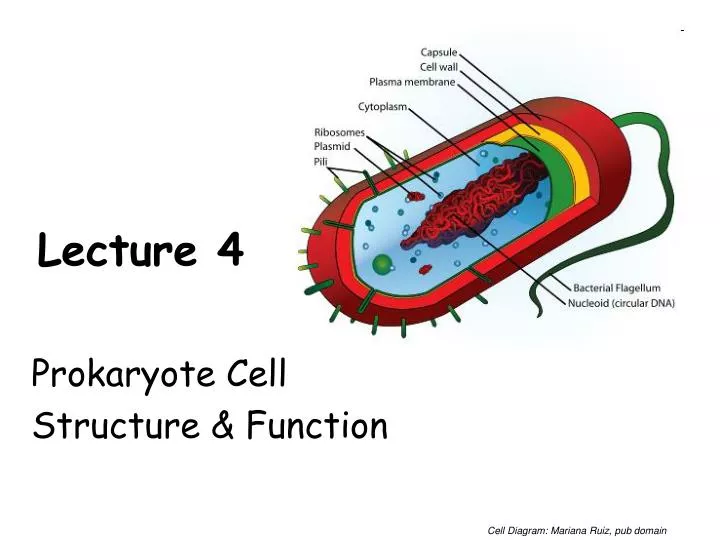
Lecture 4. Prokaryote Cell Structure & Function. Cell Diagram: Mariana Ruiz, pub domain. Size of Living Things. 1 m = 100 cm = 1,000mm = 1,000,000 µ m = 1,000,000,000nm 1mm = 1000 µ m = 1000000nm 1 µ m = 1000nm. Diagrams: http://www.cellsalive.com/howbig.htm.

E N D
Lecture 4 Prokaryote Cell Structure & Function Cell Diagram: Mariana Ruiz, pub domain
Size of Living Things 1 m = 100 cm = 1,000mm = 1,000,000 µm = 1,000,000,000nm 1mm = 1000 µm = 1000000nm 1 µm = 1000nm Diagrams: http://www.cellsalive.com/howbig.htm
The Cellular Level of Organization Living things are constructed of cells. Living things may be unicellular or multicellular. Cell structure is diverse but all cells share common characteristics. Cells are small so they can exchange materials with their surroundings. Surface area relative to the volume decreases as size of cell increases. - limits the size of cells ___________________ states: 1. All organisms are composed of one or more cells. 2. Cells are the basic unit of structure and function in organisms. 3. All cells come only from other cells.
Two basic types of cells _____________________ _____________________ Diagrams: Prokaryotic & Eukaryotic Cell, Mariana Ruiz
Prokaryotes Binary Fission Tell me about Prokaryotes… Diagrams: Prokaryotic Mariana Ruiz Binary Fission, JW Schmidt
Prokaryote Genetics ___________ Region of cytoplasm where chromosomal DNA is located. Usually a singular, circular chromosome ____________ Small extra piece of chromosome/genetic material. 5 - 100 genes Not critical to everyday functions. Can provide genetic information to promote: - Antibiotic resistance - Virulence factors (molecules produced by pathogen that specifically influence host's function to allow the pathogen to thrive) - Promote conjugation (transfer of genetic material between bacteria through cell-to-cell contact) STRUCTURE OF MICOBIAL CELLS
Prokaryotes ______________ Also known as proto-plasm. Gel-like matrix of water, enzymes, nutrients, wastes, and gases and contains cell structures. Location of growth, metabolism, and replication. ______________ Bacteria’s way of storing nutrients Staining of some granules aids in identification STRUCTURE OF MICOBIAL CELLS
Prokaryotes _______________ Found within cytoplasm or attached to plasma membrane. What are they made of? What do they do? Composed of a small (30S) subunit and a large (50S) subunit. Cell may contain thousands of ribosomes. _________________ Cellular "scaffolding" or "skeleton" within the cytoplasm. Major advance in prokaryotic cell biology in the last decade has been discovery of the prokaryotic cytoskeleton. Previously thought to be a feature only of eukaryotic cells. Eukaryotic Cells Diagrams/Photos: Fluorescent Cell: NIH, Pub Domain
Prokaryotes- Plasma Membrane Separates the cell from its environment. Phospholipid molecules oriented so that hydrophilic (__________) heads directed outward and hydrophobic (__________) tails directed inward. Proteins embedded in two layers of lipids (lipid bilayer). ___________________ to allow substances to pass into and out of the cell. Diagrams: Prokaryotic Cell, Mariana Ruiz Membrane: NIST
Prokaryotes – Plasma Membrane as a Barrier Primary function of plasma membrane →regulate movement of molecules entering or leaving cell. Movement of molecules across plasma membrane requires energy. PASSIVE TRANSPORT Movement of molecules is passive if no energy sources of the cell are expended. _____________ = when molecules move from a higher to a lower concentration. What type of things might affect the rate of diffusion? Diffusion Animation: biologycorner.com
Prokaryotes – Plasma Membrane as a Barrier PASSIVE TRANSPORT (Continued) _____________ Diffusion Proteins assist in diffusion of molecules across plasma membrane. Movement only occurs in the presence of a concentration gradient. Some molecules move across the membrane more quickly if diffusion is facilitated by a carrier molecule. Diagram: Facilitated diffusion, Mariana Ruiz
Prokaryotes – Plasma Membrane as a Barrier Osmosis Diffusion of ________ across the plasma membrane. Environment surrounding cells may contain amounts of dissolved substances (solutes) that are… equal to less than greater than …those found within the cell. Tonicity __________: no net movement of water between cell and environment __________: a higher concentration of solute. __________: a lower concentration of solute. Water will always move toward a hypertonic environment!! Diagrams: Osmosis - www.scienceaid.co.uk/biology/plants/osmosis.html Blood Cells: Mariana Ruiz
Prokaryotes – Plasma Membrane as a Barrier _______________ TRANSPORT How most molecules move across the plasma membrane. Analogous to a pump moving water uphill. Types of active transport are classified by type of energy used to drive molecules across membranes. ATP Driven Active Transport Energy from adenosine triphosphate (ATP) drives substances across the plasma membrane with the aid of carrier molecules. Diagram: Source unknown
Prokaryotes – Cell Wall • Peptidoglycan is a huge polymer of interlocking chains of identical peptidoglycan monomers. • Backbone of peptidoglycan molecule composed of two derivatives of glucose: • N-acetylglucosamine (NAG) • N-acetlymuramic acid (NAM) • NAG / NAM strands are connected by interpeptide bridges. Peptidoglycan - Rigid mechanical support - Freely permeable to solutes Image: Peptindoglycan Structure: NicolasGrandjean
Prokaryotes - Cell Wall From the peptidoglycan inwards all bacteria are very similar. Going further out, the bacterial world divides into two major classes (plus a couple of odd types). These are: Gram-positive Gram-negative Images: PHIL Public Health Image Library
Prokaryotes - Cell Wall: Gram-Negative & Gram-Positive Image: Prokaryotic Cell, Mariana Ruiz Gram +-, Julian Onions
Why are these differences in cell wall structure so important? Images: Sources unknown
Prokaryotes - Glycocalyx Some bacteria have an additional layer outside of the cell wall called the glycocalyx. This additional layer can come in one of two forms: • Slime Layer • Capsule STRUCTURE OF MICOBIAL CELLS
Prokaryotes - Glycocalyx Some bacteria have an additional layer outside of the cell wall called the glycocalyx. This additional layer can come in one of two forms: 1. ______________________ glycoproteins loosely associated with the cell wall. Slime layers cause bacteria to adhere to solid surfaces and help prevent the cell from drying out. Streptococcus The slime layer of Gram+Streptococcus mutans allows it to accumulate on tooth enamel (yuck mouth and one of the causes of cavities). Other bacteria in the mouth become trapped in the slime and form a biofilm & eventually a buildup of plaque. Staphylococcus The slime layer of Gram+Staphylococcus allows it to thrive in the salty, hypertonic environment of the skin. Glycocalyces are not specific to Gram+ or Gram- bacteria, sometimes only some members of a certain species (strains) have a glycocalyx, whereas others don’t. STRUCTURE OF MICOBIAL CELLS
Prokaryotes - Glycocalyx 2. ___________________ polysaccharides firmly attached to the cell wall. Capsules adhere to solid surfaces and to nutrients in the environment. Adhesive power of capsules is a major factor in the initiation of some bacterial diseases. Capsule also protect bacteria from being phagocitized by cells of the hosts immune system. STRUCTURE OF PROKAROTIC CELLS
Prokaryotes - Glycocalyx Bacterial Capsule and Meningococcal Infection Meningococcal infection is caused by meningococcal bacteria (Neisseria meningitidis). Causes: • meningococcal meningitis (infection of the meninges/spinal cord) • meningococcal septicaemia (blood poisoning). Of the two forms, meningococcal septicaemia is the most dangerous. Meningococcal bacteria are the most common cause of bacterial meningitis. Approximately 5% of people who suffer from meningococcal meningitis will die. Meningococcal bacteria grow in pairs called diplococci often surrounded by a capsule coat. Over a million of these would fit on the head of a pin. http://www.meningitisuk.org/about-meningitis/bacterial-meningitis.htm STRUCTURE OF PROKAROTIC CELLS
Prokaryotes - Endospores Dormant, tough, non-reproductive structure produced by small number of bacteria. Primary function of endospores: _________________________ _________________________ Resistant to radiation, desiccation, lysozyme, temperature, starvation, and chemical disinfectants. Endospores commonly found in soil and water, where they may survive for long periods of time. A stained preparation of Bacillus subtilis showing endospores as green and the vegetative cell as red Image: Stain: Jerry Keplinger, James H. Quillen College of Medicine Procedure: Source link no longer works STRUCTURE OF PROKAROTIC CELLS
_____________________ The most significant cause of pseudomembranous colitis Severe infection of the colon, often happening after normal gut flora is eradicated by use of antibiotics. The C. difficile bacteria naturally resides in the body, but becomes overgrown. C. difficile overgrowth is harmful because the bacterium releases toxins that cause: * Bloating and constipation * Diarrhea with abdominal pain * Severe diarrhea with mucus and blood present in feces * Body aches and severe abdominal pain caused from ulcerated intestines • Treatment includes specific anticlostridial antibiotics, e.g. metronidazole or vancomycin. • News story link: http://www.thesun.co.uk/sol/homepage/news/article860499.ece STRUCTURE OF PROKAROTIC CELLS
Prokaryotes – Surface Appendages Some prokaryotes have distinct appendages that allow them to move about or adhere to solid surfaces. Consist of delicate stands of proteins. ___________ Long, thin extensions that allow some bacteria to move about freely in aqueous environments. ____________ (endoflagella) Wind around bacteria, causing movement in waves. ____________ Most Gram- bacteria have these short, fine appendages surrounding the cell. Gram+ bacteria don’t have. No role in motility. Help bacteria adhere to solid surfaces. Major factor in virulence. ____________ Tubes that are longer than fimbriae, usually shorter than flagella. Use for movement, like grappling hooks, and also use conjugation pili (singular = pilus) to transfer plasmids. Images: Flagella: Mike Jones Wiki Axial Filament: Indiana USchool of Med EColi: Pub Library of Science
Neisseria gonorrhoeae Gonorrhoeae one of the most communicable disease in the US. 125 cases per 100,000. Teens 15-19 yo 634 cases per 100,000. Young adults 20-25 460 per 100,000.