Download
1 / 32
320 likes | 513 Views
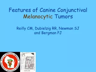
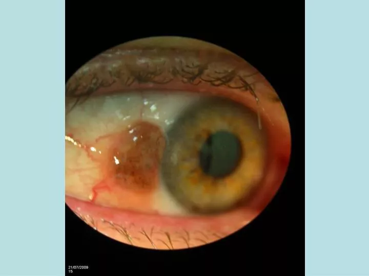
Primary conjunctival melanomas. Patient profile. 7 patients. 5 females ; 2 males. The female age range was 39-77 (median age 62). The males were aged 44 and 74. All patients had unilateral disease. 4 right eyes and 3 left eyes were affected. 14 primary invasive melanomas

E N D
Patient profile • 7 patients. 5 females ; 2 males. The female age range was 39-77 (median age 62). The males were aged 44 and 74. All patients had unilateral disease. 4 right eyes and 3 left eyes were affected.
14 primary invasive melanomas in 7 patients 3 patients Multiple mm 4 patients Solitary mm 2 juxta-limbal bulbar conjunctiva; 2 inferior fornix and inferior tarsal conjunctiva. 1 juxta-limbal bulbar, 1 juxtalimbal bulbar and non-bulbar 1 juxtalimbal bulbar and plica involvement.
Melanoma thickness • 0.1mm to 1.4 mm • pT1a to pT2b • All cases associated with in-situ MM • One case had vascular invasion.
2002 2010
19 nodules overall 7 patients 4 patients solitary 3 patients multiple 1-synchronous 2-metachronous
Location of nodules 6 patients nodules after primary Conj mm diag. 1 patient presented with nodule 19 nodules in 7 patients 11 NON-BULBAR 8 BULBAR Nodule size range 3-9mm Median-5mm
Nodules 3-102 months after first primary Conj mm (median 10m) 7 patients Systemic mets 8-37 m after First nodule 5 free of systemic mets 2 developed systemic mets Alive level 1 and 2 neck lymph nodes intra-parotid lymph node lung. Dead Bone Liver Brain
Evidence that nodules are Local METS? 2 cases Developed Systemic mets Multiple and synchronous Nodules-behaviour like mets. Well defined Cannon ball 1 nodule-necrosis Eg. Skin mm In-transits Well defined Grenz zone No overlying in-situ MM
Argument against mets. • New primaries with once-existent in-situ melanoma, with the latter regressed in response to Mitomycin C and the nodule having been ‘carved out’ Unlikely • In one case, the LCM was the presenting feature with no history of prior topical chemotherapy or surgery. • Further primary tumours developed in some cases, while on topical chemotherapy and none of these further primary tumours exhibited a well-defined, nodular morphology. • One case, the LCM developed 8 years after the primary tumour had been treated and never received MMC.
Odd distribution of LCMs? • Local factors that promote arrest and growth of the LCMs. • Surgery scarring and inflammation -damming up of tumour cells-possible but in 1 case, LCM at presentation and some cases LCM remote from surgery site. • Seeding by surgery? But 1 case presentation with LCM with no prior surgery history and no nodules at edge of dissection lines. • Dormant micromets that disseminate early…grow..? • Circulating stem cells that find niche and expand ?
All of the LCM extravascular, • Always extravascular, or whether once intravascular and have exited? • Intrinsic blood supply • Associated with a lymphocyte cap. Host reaction? LCM selected a pre-existing lymphoid niche? • LCM associated with lymphatic vessels some cases. Intraymphatic spread? Lymphangiogenesis?
Systemic mets. • 2 cases. • Is LCM a proxy measure for what is happening systemically? • Indication for sentinel LN biopsy? • Should LCMs be regarded as ‘N’ status in pathological TNM classification (like large bowel adenoca)?