Download

1 / 34
2.51k likes | 10.08k Views
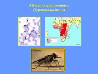
Trypanosoma. Introduction. 1- Extracellular in BLOOD & TISSUES. 2- West African Trypanosomiasis: “ West African Sleeping Sickness” caused by T. brucei gambiense. 3- East African Trypanosomiasis: “ East African Sleeping Sickness” caused by T. brucei rhodesiense.

E N D
Trypanosoma Introduction 1- Extracellular in BLOOD & TISSUES 2- West African Trypanosomiasis: “ West African Sleeping Sickness” caused by T. brucei gambiense. 3- East African Trypanosomiasis: “ East African Sleeping Sickness” caused by T. brucei rhodesiense. 4- Chronic form: caused by T. brucei gambiense. While Acute Form is caused by T. brucei rhodesiense. 5- African Sleeping Sicknessis the 3rd important parasitic disease globally after Malaria & Schistosomiasis 6- West African Sleeping Sicknessisin regions along riverside while East African Sleeping Sickness is in Forest regions (Savannas). Dr. RAAFAT T. MOHAMED
Trypanosoma Causes Trypanosomiasis West African Trypanosomiasis East African Trypanosomiasis American Trypanosomiasis T.brucei gambiense T.brucei rhodesiense T.cruzi Sleeping sickness Chagas’ disease Transmitted by Transmitted by Glossina (tsetse fly) Triatoma (winged bug) Dr. RAAFAT T. MOHAMED
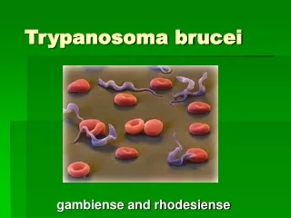
Trypanosoma Morphology Exist into 2 interchangeable forms: Trypomastigote in Blood/ Lymph / tissue space of various organs & C.N.S is terminal & fatal Epimastigote in salivary gland of vector & Culture media. Trypomastigote (Polymorphic Trypanosomes Spindle shaped – Central nucleus – free flagellum – undulating membrane. 3 forms 1- long Slender Form (30µ): active motile with free flagellum. 2- Short stumpy Form (15µ): sluggish without free flagellum. 3- Intermediate Form (20µ): with a short free flagellum. Dr. RAAFAT T. MOHAMED
Geographical Distribution of African Trypanosomiasis G.palpalis G.morsitans In West Africa In East Africa Dr. RAAFAT T. MOHAMED
Trypanosoma brucei causing Sleeping Sickness West Africa East Africa T.brucei gambiense T.brucei rhodesiense Less plentiful More plentiful Cannot live in lab animals Can live in lab animals Nucleus is shifted posteriorly Reservoir host: goats, cattle & pigs Reservoir host: wild game animals Transmitted by: G.palpalis Transmitted by: G.morsitans Dr. RAAFAT T. MOHAMED
Mechanism of disease transmission by Glossina Trypomastigotes (polymorphic trypanosomes) Diagnostic stage 12-42µ Bite of ♂& ♀ Glossina 3 weeks Salivary gland Epimastigote Full of short stumpy metacyclic Trypomastigote Infective stage Biological transmission Dr. RAAFAT T. MOHAMED
African Trypanosomiasis life cycle Life cycle of Trypanosoma brucei gambiense & T. b. rhodesiense Dr. RAAFAT T. MOHAMED
African Trypanosomiasis life cycle Dr. RAAFAT T. MOHAMED
African Trypanosomiasis life cycle Dr. RAAFAT T. MOHAMED
Pathogenesis and Clinical Picture Incubation period (2 weeks) Trypanosoma chancre (at the site of bite) Via lymphatics: enlarged lymph nodes especially posterior cervical region. (Winterbottom’s sign) Via blood stream: headache, fever(fluctuating), muscle & joint pain, irregular erythematous rash. Invasion of bone marrow (hypoplastic anaemia) Enlarged liver & spleen, generalized weakness. severe headache, mental apathy, slow speech, deep sleep, coma & death Invasion of CNS: In East African Trypanosomiasis: Disease runs more rapid & fatal course Dr. RAAFAT T. MOHAMED
Pathogenesis and Clinical Picture Winterbottom sign Trypanosoma chancre Emaciation جلد على عظم Coma before death Dr. RAAFAT T. MOHAMED
Clinical Picture Progressive disease may lead to the following C.N.S manifestations:- 1- Insomnia أرق 2- Mood changes (dullness بلاهة / apathy لامباله) 3- Motor & Sensory Disorders: (Hyperesthesia فرط الحس / slurred speech كلام متداخل / abnormal gait مشيه غير طبيعية). 4- Convulsions 5- Epilepsy داء الصرع Terminal stage: 1- Permanent Sleep. 2- 2ry Bacterial infection. 3- Coma & Death. Dr. RAAFAT T. MOHAMED
Diagnosis 1- Clinical picture 2- Demonstration of trypanosomes: Polymorphic Trypanosomes - Microscopic examination of unstained or stained blood films - Culture on suitable medium (N.N.N OR Weinmann’s media to detect Epimastigote) - Animal inoculation N.B. in case of T.brucei rhodesiense injected in lab Animal produce a new form “Posterior Nucleus Shift” Dr. RAAFAT T. MOHAMED
Diagnosis C.S.F Dr. RAAFAT T. MOHAMED
Diagnosis 3- Serological test: Increased total IgM level in serum due to antigenic variationof the surface coat of the parasite. Trypanosome posses genes that code for about1000variant formsof their surface glycoproteins (SVG). Switch to a different variant produces a new generation not susceptible to attack by immune factors specific to the previous generation. Trypanosomes can evade(تهرب) the immune system زيادة مطردهWhy in African Trypanosomiasis infection, there is a sustained elevated IgM level ??????? Dr. RAAFAT T. MOHAMED
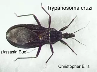
Trypanosoma cruzi causing Chagas’ disease Winged bug Prominent kinetoplast Kissing bug Triatoma or Rhodnius Trypanosoma cruzi C-shaped Dr. RAAFAT T. MOHAMED
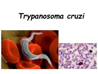
Trypanosoma cruzi Morphology Trypomastigote (Monomorphic) Slender shaped (20µ) – Central nucleus – C or U-shaped –Free flagellum 1/3 body- Large bulging peripheral kinetoplast Amastigote Obligatory intracellular – mainly in cardiac & Skeletal muscles – Brain meninges – Nerve ganglia – cells of GIT …. etc Epimastigote (Vector only) Spindle shape– Kinetoplast anterior to central nucleus– Undulating membrane is short – terminal free flagellum Dr. RAAFAT T. MOHAMED
Mechanism of disease transmission by winged bug T.cruzi in human blood Cyclopropagative transmission Alimentary canal of bug Short stumpy metacyclic trypomastigote (infective stage) Epimastigote form Pass out with faeces Dr. RAAFAT T. MOHAMED
AMERICAN TRYPANOSOMIASIS LIFE CYCLE OF Trypanosoma cruzi Dr. RAAFAT T. MOHAMED
Mode of infection Mainly by Contamination of skin abrasion (خدوش بالجلد) by winged bug faeces Cone nose Bug – kissing Bug –Assassin bug Rarely by Through infected blood transfusion Through infected mother’s milk Through the placenta Dr. RAAFAT T. MOHAMED
Life cycle of Trypanosoma cruzi Dr. RAAFAT T. MOHAMED
Life cycle of Trypanosoma cruzi Dr. RAAFAT T. MOHAMED
Pathogenesis and Clinical Picture I- Acute Form Chagoma occurs at the site of bite. Parasite reaches regional lymph nodes To Blood To Organs and tissues Fever, enlarged lymph nodes, skin rash, enlarged liver & spleen. Romana’s sign(Unilateral conjunctivitis appear suddenly together with oedema of upper & lower eye lids & cheek) Meningoencephalitis, heart failure Death or pass to Chronic form Dr. RAAFAT T. MOHAMED
Pathogenesis and Clinical Picture II- Chronic form Parasite produces antigens similar to patient’s self antigens: Amastigote form of T.cruzi The body produces auto-antibodies that cause damage to: • Heart muscle fibres: congestive heart failure. • Oesophageal muscle fibres: عصر البلع megaoesophagus and dysphagia. Destruction of Auerbach’s plexus • Colon muscle fibres: megacolon and constipation. • CNS or thyroid gland Exacerbation of infection in immunosuppressed patients. Dr. RAAFAT T. MOHAMED
Diagnosis Finding the parasite in: Blood film (C-shaped T.cruzi) Biopsy from lymph node, liver or spleen (amastigotes) Culture(Epimastigotes) Xenodiagnosis Serological tests Cruzin test (I.D.) Molecular techniques Dr. RAAFAT T. MOHAMED
Diagnosis (Xenodiagnosis) Highly efficient – demonstrate low level of parasite in blood Method: A Laboratory bred winged bug is starved for 2 weeks then fed on suspected patient’s blood – 30 days later, it faeces & gut examined for trypanosomes. Dr. RAAFAT T. MOHAMED
Diagnosis Winged Bug Trypomastigote Amastigote Chagoma Romana’s sign Dr. RAAFAT T. MOHAMED
In early stage of the disease: Pentamidine OR Suramin In late stages of the disease: Tryparsamide For both early and late stages of the disease: Eflornithine (DFMO) Ornidyl Nifurtimox inhibits intracellular development . Drug of choice in acute and early chronic OR Primaquine destroys Trypanosoma in blood Treatment Sleeping Sickness Chagas Disease Dr. RAAFAT T. MOHAMED
Treatment of patients Control of vectors (Glossina) Pentamidine as prophylactic drug Treatment of patients Control of vectors (Triatoma) Elimination of reservoir hosts Control Chagas’ disease Sleeping Sickness Dr. RAAFAT T. MOHAMED
M.C.Q. Protozoal infections that may cause fever and hepatosplenomegaly a- Visceral leishmaniasis b- African trypanosomiasis (sleeping sickness) c- American trypanosomiasis (Chagas’ disease) d- Non of the above e- All of the above Smear taken from the edge of oriental sore reveals: a- promastigote c- amastigote d- trypomastigote b- epimastigote Dr. RAAFAT T. MOHAMED
M.C.Q. Protozoa causing conjunctivitis include: a- Trypanosoma cruzi b- T.brucei gambiense c- T.brucei rhodesiense d- Leishmania donovani Winterbottom’s sign is seen in: a- Cutaneous leishmaniasis b- Visceral leishmaniasis c- African trypanosomiasis d- American trypanosomiasis Dr. RAAFAT T. MOHAMED
M.C.Q. In African trypanosomiasis, the infective stage is found in: Short stumpy metacyclic trypanosomes a- Saliva of Triatoma c- Stool of Triatoma d- Stool of Glossina b- Saliva of Glossina Posterior nuclear shift occurs in: a- Trypanosoma cruzi b- Trypanosoma gambiense c- Trypanosoma rhodesiense d- Trichomonas vaginalis Dr. RAAFAT T. MOHAMED
M.C.Q. In chronic Chagas’ disease, the main lesions are in: a- Digestive and respiratory tracts. b- Heart and liver. c- Heart and digestive tract. d- Liver and spleen. Megacolon associated with Chagas’ disease: a- Is manifested by diarrhoea. b- Occurs early in the disease. c- Is due to oedema of the mucosa. d- Is associated with constipation. Dr. RAAFAT T. MOHAMED
Compare between Romana’s sign Acanthamoeba affection of the eye Inflammation of the conjunctiva Inflammation of the cornea Ulceration Perforation Ocular pain & affection of vision Mode of infection Contamination of skin abrasions by winged bug (Triatoma) faeces Occurs through corneal trauma Exposure to contaminated water Wearing contaminated contact lenses Short stumpy metacyclic trypanosomes Dr. RAAFAT T. MOHAMED