Download
1 / 108
1.09k likes | 1.14k Views
Saladin Ch. 21. Lymphatic & Immune Systems. Lymphatic System General. Lymphatic System - composed of lymph, lymphatic vessels, and lymphatic tissue Functions of Lymphatic system functions Draining of interstitial fluid

E N D
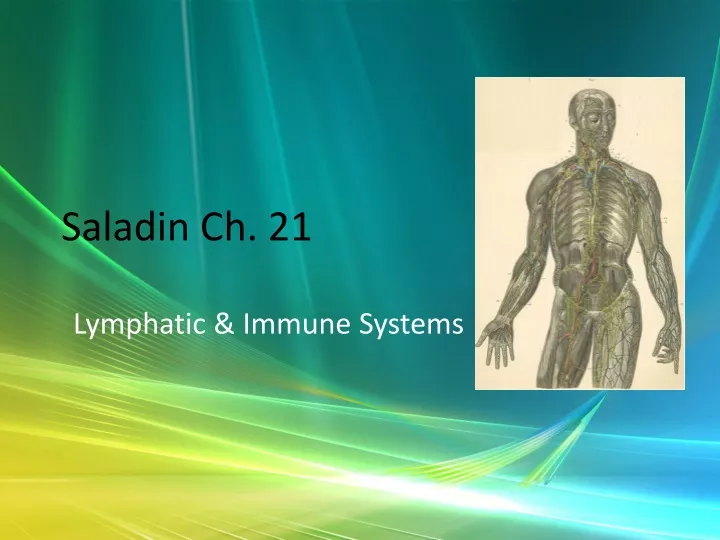
Saladin Ch. 21 Lymphatic & Immune Systems
Lymphatic System General • Lymphatic System - composed of lymph, lymphatic vessels, and lymphatic tissue • Functions of Lymphatic system functions • Draining of interstitial fluid • Transporting dietary lipids/lipid soluble vitamins from GI to blood. • Facilitating the Immune Response - by B and T lymphocytes.
Lymph & Lymphatic Vessels Lymph • Usually clear, colorless, derived from blood plasma but with less protein • Lacteals - specialized capillaries along the small intestine - pick up lipids - gives fluid white, opaque look = chyle • Contains large numbers of lymphocytes, etc.
Lymph & Lymphatic Vessels Lymphatic capillaries • Closed ends; One way flow - wall cells overlap; fluid pushes in & can separate the cells. • Anchoring filaments - hold capillaries in place & help open gaps when interstitial fluid builds up. • Location - not in cartilage, epidermis, CNS, parts of spleen or red marrow.
Lymph & Lymphatic Vessels Lymph trunk and ducts • In embryo form from buds from veins - similar structure. • Flow - capillaries into vessels into nodes. After passing through a string of nodes - goes into "trunks“
Lymph & Lymphatic Vessels • [Trunks: Lumbar, intestinal, subclavian bronchomediastinal, & juglar] • From trunks, lymph flows into either the thoracic duct or the right lymphatic duct • From the ducts, flow is into venous blood
Lymph & Lymphatic Vessels • Right lymphatic duct • Drains from upper R side • Drains into R. subclavian vein • Trunk feeders - R juglar from R head and neck, R subclavian from R upper limb, R bronchmediastinal from R thorax.
Lymph & Lymphatic Vessels • Thoracic Duct - begins as cisterna chyli - anterior to L2 vertebra • Receives from left of head & neck, chest, left upper limb, & all of the body below the ribs. • Drains into the left subclavian vein • Feeder trunks - lower body -R & L lumbar, intestinal
Lymph & Lymphatic Vessels • Lumbars drain from lower limbs, pelvis, kidneys, adrenals & abdominal wall. • Intestinal drains from intestines, pancreas, spleen & liver • From the neck, the thoracic duct gets lymph from the l. juglar, l. subclavian, l. bronchomediastinal trunks
Lymphatic Cells • Lymphocytes - stem cells divide to produce B & T cells • Produced in red marrow • B cells may become plasma cells that produce antibody • Pluripotent cells in red marrow pre-T cells thymus to mature.
Lymphatic Cells • Macrophages [Phago. & APC's] • Dendritic Cells [activate T cells - APC's] • Reticular cells – fibroblasts that produce reticular fibers for tissue “stroma”
Lymphatic Tissue • Aggregations of lymphocytes in connective tissues of mucus membranes • MALT, Galt, nodules, Peyer's patches
Lymphoid Organs • Include marrow, thymus, lymph nodes, spleen, tonsils • Red Bone Marrow --> lymphocytes [review blood chapter]
Lymphoid Organs • Thymus Gland • Functions in T cell maturation – Reticular epithelial cells produce thymic hormones that aid in maturation of T cells • Below sternum - large in infants, shrinks with age from puberty on. • 2 lobes – have capsule with trabeculae
Lymphoid Organs • Lobules - 2 regions - cortex and medulla • Cortex - deep staining - lymphocytes & reticular epithelial cells • Medulla - paler - mostly reticular epithelium - fewer lymphocytes -Has thymic corpuscles [Hassall's] - concentric whorls of flattened reticular epithelium
Lymphoid Organs • Lymph Nodes - about 600 bean-shaped organs • Concentrated in axillae, groin & near mammaries. • Function: • Trap material on reticular fibers, • Clear out foreign & damaged materials through phagocytosis, • Immune processes
Lymphoid Organs • Structure - stroma & parenchyma • Stroma - capsule - dense connective tissue covering - has trabeculae.Supports & holds vessels. Also has network of reticular fibers & fibroblasts.
Lymphoid Organs • Parenchyma - 2 regions - cortex and medulla • Cortex - outer and inner • Outer - lymphatic nodules of B cells. Germinal centers - where B cells proliferate into plasma cells. Dendritic cells - APC's - initiate immune response. Also have macrophages. • Inner - T-cells
Lymphoid Organs • Medulla – B & T cells, plasma cells tightly packed in "medullary cords“ • Lymph nodes have unidirectional flow - enters through afferent vessels sinuses efferent lymphatic vessels. [Hilus - place where efferent vessels emerge.] • Lymph flows through a series of nodes --> quite clean at the end.
Lymphoid Organs • Metastasis - spread of cancer. • Sites for metastasis are predictable based on lymphatic flow patterns. • Conversely, when a secondary tumor is found, the primary can usually be located by going in reverse back the flow paths.
Lymphoid Organs • Tonsils • Pharyngeal = adenoid, palatine =at base of palatine bones, lingual = base of tongue, tubal = around auditory tube openings • No capsule – “crypts” that trap bacteria & particulate material
Lymphoid Organs • Spleen - largest lymphatic structure - between stomach & diaphragm. • Function: • Immunologic – precipitate, kill antigens • Clean up old rbc’s, • Store platelets, • Stabilize blood volume
Lymphoid Organs • Parenchyma • White pulp - Lymphocytes & macrophages • Red pulp – venous sinuses filled with blood & Billroth’s [splenic] cords [rbc’s, macrophages, lymphocytes, B cells, granulocytes pressed together]
Lymphoid Organs • Stroma • Capsule with trabeculae covered, with serous visceral peritoneum. • Interior reticular tissue.
Non-specific Disease Resistance • Non-specific Resistance to Disease –> immediate protection against a wide variety of pathogens & foreign substances • NOmemory – always the same • Pathogen = a disease-causing agent
Non-specific Disease Resistance • Lines of Defense: 1 - external barriers - non-specific 2 - non-specific internal responses 3 - immune system - specific
Non-specific Disease Resistance • External Barriers Skin & Mucous Membranes • Mechanical factors – physical barriers – intact skin, tight junctions • Mucous membranes – physical barriers also – mucous catches dirt, etc.; hairs filter out material, cilia sweeps out invaders
Non-specific Disease Resistance • Chemical Factors: • Defensins • pH of skin [3-5] antimicrobial • Sweat – flushes, contains lactic acid = acid mantle
Non-specific Disease Resistance • Tears – dilute agents. Also some lysozyme - antibiotic properties • Saliva – same Urine / vaginal secretions • Dermal hyaluronic acid - viscous - hard to traverse Some organisms have hyaluronidase to dissolve it.
Non-specific Disease Resistance • Internal Defenses - Leukocytes & Macrophages Phagocytes – eat foreign matter • Neutrophils – in most body tissues. In addition to phagocytosis, use respiratory burst - series of reactions/agents that create H2O2, HClO & superoxide ions that destroy bacteria
Non-specific Disease Resistance • Eosinophils – can attack parasitic worms, promote basophil action, reduce inflammatory response • Basophils - secrete histamine - vasodilator and Heparin - anticoagulant [both also released by mast cells].
Non-specific Disease Resistance • Lymphocytes - 3 classes [specific and non] 80% T, 15% B, 5% NK • Monocytes • Special "macrophages" - dendritic cells, microglia, alveolar, hepatic
Non-specific Disease Resistance • Internal Defenses - Antimicrobial Proteins • Interferons – produced by virally infected lymphocytes, macrophages, & fibroblasts • Attach to uninfected cells & induce synthesis of proteins that interfere with viral replication • Protects these cells from infection • Also activate NK cells
Non-specific Disease Resistance • Complement – a group of 30+ proteins that are synthesized by the liver & in the blood normally inactivated • When activated – enhance all parts of the immune response as well as allergic responses • Activated 3 ways - complement fixation [Ab], alternative path [ spontaneous - no Ab], Lectin path - attach to sugars on microbes
Non-specific Disease Resistance • 4 methods. • Inflammation - C3a stimulates mast and basophils to release histamine and initiate the IR, activates & attracts neutrophils • Immune Clearance - C3b - binds Ab-Ag complexes to rbc's - collected by macrophages in liver & spleen
Non-specific Disease Resistance • Phagocytosis - opsonization by C3b • Cytolysis - C3b initiated.--> cascade -->makes a hole in membrane of target cell
Non-specific Disease Resistance • Immune Surveillance • NK cells patrol for foreign agents and diseased host cells - destroy them • Use perforins + granzymes