Download
1 / 5
50 likes | 310 Views
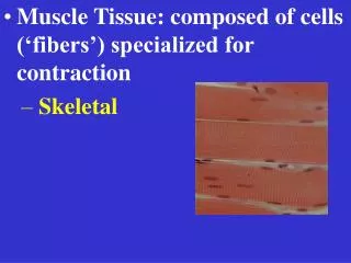
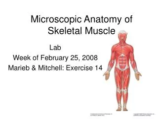
Microscopic Anatomy of Skeletal Muscle Fibers. Long cylindrical cells Multiple nuclei just below sarcolemma Glycosomes- granules of stored glycogen Myoglobin- red pigment stores O 2 Myofibrils- rodlike structures that run parallel to length of cell, densely packed

E N D
Microscopic Anatomy of Skeletal Muscle Fibers • Long cylindrical cells • Multiple nuclei just below sarcolemma • Glycosomes- granules of stored glycogen • Myoglobin- red pigment stores O2 • Myofibrils- rodlike structures that run parallel to length of cell, densely packed • Made of myofilaments- contractile elements of skeletal muscles
Myofilaments • Thick filaments- myosin • rodlike tail, ends in flexible hinge with 2 globular heads • Form cross bridges with thin filaments • Contain actin binding sites and ATPase enzymes • Thin filaments- actin • subunits are globular= G actin • Tropomyosin- strands run along actin, help stiffen actin molecule, in a relaxed muscle cell they block myosin binding sites • Troponin- 3 polypeptide complex, 1= binds to actin to inhibit contractions, 2= binds to tropomyosin and helps position actin, 3= Ca2+ binding site
Striations and Sarcomeres • Striations- repeating series of dark A band & light I bands • H zone- lighter stripe in midsection of A band • M line- dark line that bisects H zone vertically • Z disc- dark midline interruption of I bands • Sarcomere- region of myofibril between 2 sucessive Z discs, smallest functional unit of muscle fiber
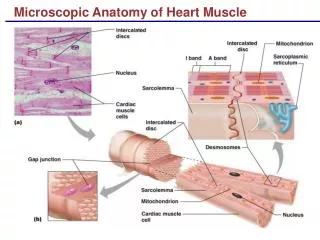
Microscopic Anatomy Cont’d • Sarcoplasmic reticulum- elaborate SER, interconnecting tubules surround each myofibril • most run longitudinal but some larger channels cross perpendicular at A band- I band junctions= terminal cisternae • T tubules- at each A/I band junction, sarcolemma of muscle cell penetrates into cell interior= elongated tube • can/do conduct impulses to deep regions of muscle cell and to every sacromere, ensure that every myofibril contracts at same time
Sliding Filament Theory • “during contraction, thin filaments slide past thick ones so that the actin and myosin filaments overlap to a greater degree” • Shortening occurs when tension generated by cross bridges exceeds forces opposing shortening, contraction ends when cross bridges become inactive, tension declines Sarcomere Shortening (499.0K) Actin Myosin Bridge