Download
1 / 8
90 likes | 591 Views
Microscopic Anatomy of Skeletal Muscle. Lab Week of February 25, 2008 Marieb & Mitchell: Exercise 14. Muscle Fibers. multinucleate nuclei in peripheral area myofibrils I and A bands myofilaments actin & myosin. Sarcomere. Z to Z M line in middle

E N D
Microscopic Anatomy of Skeletal Muscle Lab Week of February 25, 2008 Marieb & Mitchell: Exercise 14
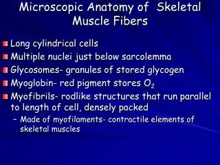
Muscle Fibers • multinucleate • nuclei in peripheral area • myofibrils • I and A bands • myofilaments • actin & myosin
Sarcomere • Z to Z • M line in middle • thick and thin filaments overlap and interact to contract the myofibril
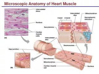
SR, T tubules and Triads • At the junction of A and I, the sarcolemma indents to form the T tubule • Sarcoplasmic Reticulum (SR) like smooth ER in most cells • terminal cisternae: SR next to T tubules • Triads (terminal cisternae – T tubule – terminal cisternae)
Fiber to Muscle Organization • muscle fiber • endomysium • perimysium • fascicle • epimysium • skeletal muscle • deep fascia • tendons or aponeuroses
Activity 1 • Model of Muscle Cell • Look at fresh chicken muscle under microscope • Look at slide with cross section of skeletal muscle and locate the structures in Fig 14.4 (if visible) Activity 2
The Neuromuscular Junction • axon • axon terminal • motor unit • 1 neuron and all the muscles it stimulates • Plate 4, pg 693
Activity 3 • Model of skeletal muscle cell with NMJ? • Slide with motor unit • axon fibers • axon terminal • COMPARE WITH PLATE 4 on pg 693