Download
1 / 52
530 likes | 574 Views
Discover the intricacies of the lymphatic system, its vessels, cells, and organs with a focus on the movement, filtration, and immune response essential for maintaining overall health. Learn about lymphatic capillaries, trunks, lymphoid cells, and tissues, including lymph nodes and organs like the spleen, thymus, and tonsils. Explore the mechanisms of lymph transport, immune system activation, and the causes of edema associated with lymphatic system dysfunction.

E N D
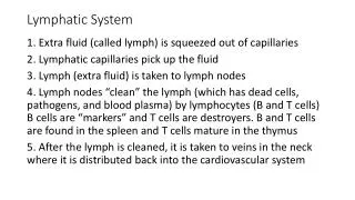
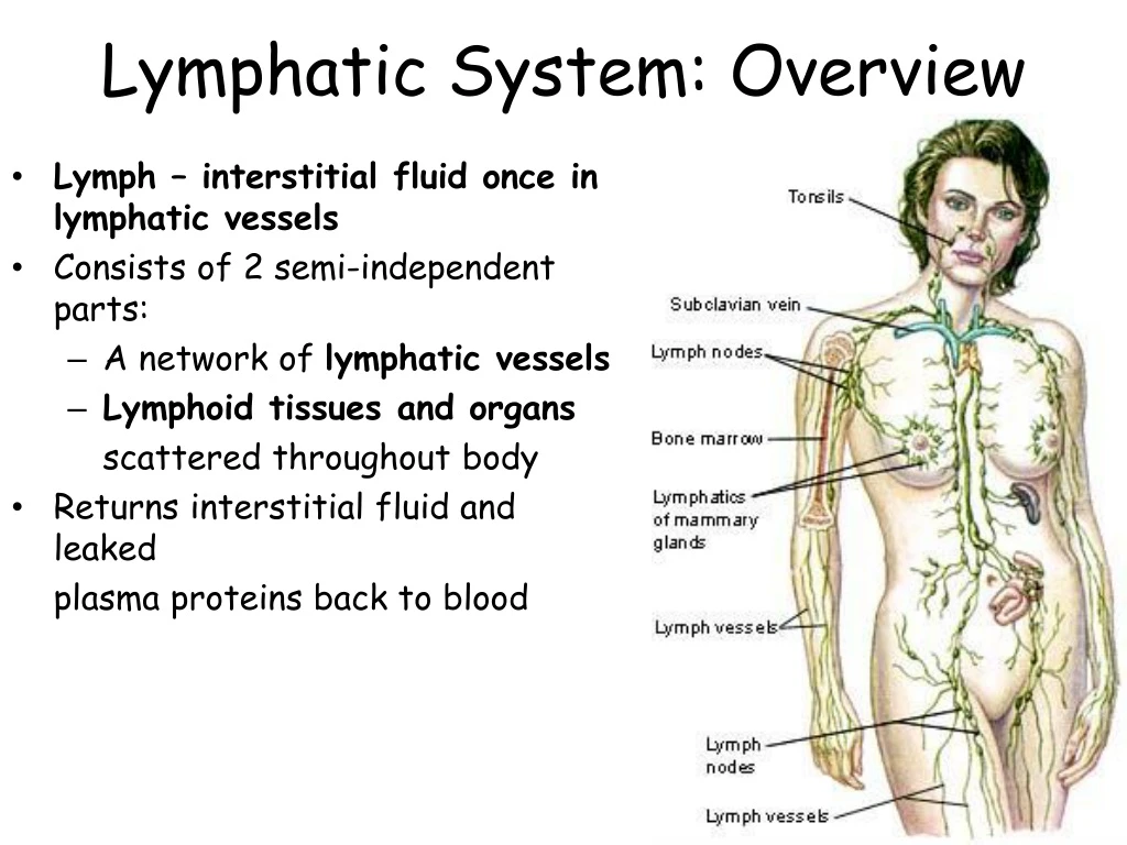
Lymphatic System: Overview • Lymph – interstitial fluid once in lymphatic vessels • Consists of 2 semi-independent parts: • A network of lymphatic vessels • Lymphoid tissues and organs scattered throughout body • Returns interstitial fluid and leaked plasma proteins back to blood
One-way system: lymph flows toward the heart • Lymph vessels include: • Microscopic, permeable lymphatic capillaries • Lymphaticcollecting vessels • Lymphatic trunks and ducts
Lymphatic Capillaries • Similar to blood capillaries, with modifications: • More permeable • Loosely joined minivalves • Withstand interstitial pressure and remain open • Minivalves function as one-way gates that: • Allow interstitial fluid to enter, but not escape lymph capillaries • During inflammation, lymph capillaries absorb cell debris, pathogens, cancer cells • Lacteals – specialized lymph capillaries present in intestinal mucosa - absorb digested fat and deliver chyle to the blood
Lymphatic Vessels & Transport • Have 3 tunics (as veins) • Have thinner walls, with more internal valves • Collecting vessels in the skin travel along superficial veins • Deep vessels travel along arteries • Nutrients are supplied from branching vasa vasorum (network of small arterioles, capillaries, and venules that supply the outer tissue of large blood vessels) • The lymphatic system lacks a pumping organ • Vessels are low-pressure conduits • Uses the same methods as veins to propel lymph: • Pulsations of nearby arteries • Contractions of smooth muscle in the walls of lymphatics Lymphatics & the Breast
Lymphatic Trunks • Lymphatic trunks are formed by union of largest collecting ducts • Major trunks include: • Paired lumbar, bronchomediastinal, subclavian, and jugular trunks • A single intestinal trunk • Lymph is delivered into 1 of 2 large trunks • Right lymphatic duct – drains right upper arm and the right side of head and thorax • Thoracic duct – arises from cisterna chyli and drains rest of body
Lymphoid Cells • Lymphocytes are THE main cells involved in immune response • 2 main kinds: T cells and B cells, they protect body against antigens • Antigen – anything the body perceives as foreign: bacteria, viruses, mismatched RBCs or cancer cells • T cells: manage immune response; attack foreign cells • B cells: produce plasma cells, which secrete antibodies => Antibodies immobilize antigens Other lymphoid cells: • Macrophages – phagocytize foreign substances and activate T cells • Dendritic cells – spiny-looking cells, functions similar to macrophages • Reticular cells – supports other cell types in lymphoid organs
Lymphoid Tissue Lymph Nodes • Scattered reticular tissue elements in every body organ • Principal lymphoid organs of the body • Within connective tissue and along lymphatic vessels • Aggregations of nodes occur near body surface in inguinal, axillary, and cervical regions of the body • 2 basic functions: • Filtration – macrophages destroy microorganisms and debris • Immune system activation – monitor and attack antigens
Structure of a Lymph Node • Nodes are bean-shaped and surrounded by a capsule • Trabeculae extends inward from capsule and divide node into compartments • Nodes have 2 histologically distinct regions: a cortex and a medulla • Cortex contains follicles with dividing B cells • Cortex houses T cells
Lymph nodes 2 Regions: Cortex B cells T cells Medulla Lymph Sinuses & macrophages Lymph Nodes Animation
Lymphoid Organs Lymph Nodes Spleen Thymus Tonsils Peyer’s patches MALT
Spleen White pulp B and T cells carry out immune function. Red pulp Removes aged and defective RBCs Stores breakdown products of RBCs Erythrocyte production in fetus Stores blood platelets Has regenerative properties
Thymus Secretes thymopoietin, thmosins to make T-cells immunocompetent Bilobed organ Trabeculae divide lobe into lobules. Thymic corpuscles
Tonsils & Adenoids Trap bacteria which work their way into the follicles where they are destroyed This helps develop memory
Appendix • Possibly works with the Peyer's patches to help defend against invaders from the digestive system
Aggregates of Lymphoid Follicles • Peyer’s patches – isolated clusters, similar to tonsils • In the wall of the distal portion of the small intestine • Similar structures are found in the appendix • Peyer’s patches and the appendix: • Destroy bacteria • Generate “memory” lymphocytes for long-term immunity
MALT Mucosa-associated lymphatic tissue Peyer’s patches, tonsils, and appendix (digestive tract) Lymphoid nodules in the walls of the bronchi (respiratory tract) MALT protects the digestive and respiratory systems from foreign matter
Causes of Edema Edema Accumulation of interstitial fluid Blockage of lymphatic system Increased pressure in veins Lack of albumin Decreases fluid returning to blood capillaries by osmosis Inflammation
Homeoimbalances of the Lymphatic System • Autoimmune Lymphoproliferative Syndrome (ALPS) • Lymphatic Filariasis • Mesenteric Lymphadenitis • Swollen Lymph Nodes • Castleman Disease • Adenoids • Splenomegaly • Hodgkin's disease • Kawasaki disease
Immunity: 2 Defense Systems • Innate (nonspecific) system responds quickly and consists of: • First line of defense – skin and mucosae prevent entry of microorganisms • Second line of defense – antimicrobial proteins, phagocytes • Inhibit spread of invaders throughout the body • Inflammation is its most important mechanism • Adaptive (specific) defense system • Third line of defense – mounts attack against foreign substances • Has memory, antigen-specific, and antigen-mediated immunity • Works in conjunction with the innate system • Recognizes specific foreign substances • Immobilizes, neutralizes, or destroys foreign substances
First line of defense: Surface membrane barriers • Skin and mucous membrane • Layered epidermis and shedding of epithelial cells • Sebum inhibits growth of bacteria and fungi • Mucous traps microbes, dust and pollutants. • Lacrimal apparatus • Saliva • Vaginal secretions • Flow of urine • Defecation and vomiting • Gastric juices destroy bacteria and their toxins
First line of defense: Surface membrane barriers • Skin and mucous membrane • Layered epidermis and shedding of epithelial cells • Sebum inhibits growth of bacteria and fungi • Mucous traps microbes, dust and pollutants. • Lacrimal apparatus • Saliva • Vaginal secretions • Flow of urine • Defecation and vomiting • Gastric juices destroy bacteria and their toxins
Second line of defense: chemical and cellular defenses • Antimicrobial proteins • Interferon • Complement • Transferrins • Natural killer cells • Phagocytes • Neutrophils • Dendritic cells • Macrophages • Wandering • Fixed • Eosinophils
Interferons • Produced by lymphocytes, macrophages and fibroblasts. • Interfere with translation of viral proteins • Degrade viral RNA • Activate macrophages and NK cells • Interferon Animation
Complement Complement Cascade Animation
Phagocytes • Macrophages are the chief phagocytic cells • Free macrophages wander in search of cellular debris • Kupffer cells (liver) and microglia (brain) are “fixed” macrophages • Neutrophils become phagocytic when encountering infectious material • Eosinophils are weakly phagocytic against parasitic worms • Mast cells bind and ingest a wide range of bacteria Mechanism • Pseudopods engulf the antigen into a phagosome • Invaders are digested by proteolytic enzymes • Indigestible and residual material is removed by exocytosis Figure 21.2a
Fever • Abnormally high body temperature in response to invading microorganisms • Body’s thermostat is reset upwards in response to pyrogens, chemicals secreted by leukocytes and macrophages exposed to bacteria and other foreign substances • High fevers are dangerous because they can denature enzymes • Moderate fever can be beneficial, as it causes: • Liver and spleen to sequester iron and zinc • Increases metabolic rate, which speeds up tissue repair
Inflammatory response Stages Inflammation Animation • Release of Chemical Alarms • Vasodilatation & Permeability of BV • Emigration of phagocytes: Dispose cellular debris & pathogens • Sets the stage for repair • Prevent spread of damaging chemicals & pathogens Signs of inflammation • Redness • Heat • Swelling • Pain • Impairment of function
Adaptive Resistance • Specificity—recognition of particular antigens • Memory—remembers previously encountered antigens • Systemic—immunity is not restricted to the initial infection site • Immune responses • Antibody-mediated or humoral immune responses (late 1800s) • Cell-mediated immune responses (mid 1900s)
Antigens and antigen receptors • Antigens can be entire microbes, parts of microbes or chemical components of pollen, egg white, blood cells,…….
Antibodies “immunoglobulins” • Four looping polypeptide chains linked together through disulfide bonds. • Heavy chains are identical and have a hinge • Light chains are half as long. • Variable region is the antigen binding site • Constant region forms the stem of the antibody and determines its class • Do not destroy antigen; inactivate and tag it for destruction • form an antigen-antibody (immune) complex
Antibody Action • Defensive mechanisms used by antibodies: • Complement fixation – antibodies bound to cells change shape and expose complement binding sites • Complement activation – uses a positive feedback cycle to promote phagocytosis • Neutralization – antibodies block binding sites on viruses • Precipitation – soluble molecules are cross-linked into large insoluble complexes
Immunoglobulin classes • IgD is attached to B-cell plasma membrane • IgM is released during primary response. Indicates current infection. • IgG is the most aboundant. Can cross placenta & blood vessel walls. • IgA found in body secretions prevents attachment to body surfaces. • IgE causes release of histamine (allergies) by attaching to mast cells & basophils.
Immunological memory • Primary immune response • Secondary immune response
Lymphocytes • Immature lymphocytes released from bone marrow are essentially identical • Whether a lymphocyte matures into a B cell or a T cell depends on where in the body it becomes immunocompetent • B cells mature in the bone marrow • T cells mature in the thymus
Key: Red bone marrow = Site of lymphocyte origin = Site of development of immunocompetence as B or T cells; primary lymphoid organs = Site of antigen challenge, activation, and final diff erentiation of B and T cells Immature lymphocytes Circulation in blood 1 1 Lymphocytes destined to become T cells migrate to the thymus and develop immunocompetence there. B cells develop immunocompetence in red bone marrow. 1 Thymus Bone marrow 2 Immunocompetent, but still naive, lymphocyte migrates via blood 2 2 After leaving the thymus or bone marrow as naïve immunocompetent cells, lymphocytes “seed” the lymph nodes, spleen, and other lymphoid tissues where the antigen challenge occurs. Lymph nodes, spleen, and other lymphoid tissues 3 3 Antigen-activated immunocompetent lymphocytes circulate continuously in the bloodstream and lymph and throughout the lymphoid organs of the body. 3 Activated Immunocompetent B and T cells recirculate in blood and lymph Figure 20.8
T Lymphocytes • CD4 T cell - also known as a T Helper (Th) cell • CD8 T cell - also known as a Cytotoxic T (Tc) cell
B Lymphocytes • Clonal Selection • Production of clones initiated by antigen binding • Plasma cells secrete antibodies • Memory cells are long lived
Humoral & Cell-Mediated Immunity • Humoral = antibody mediated immunity • Involves B cells • Antibodies circulate through “humors” inactive and mark invaders for destruction • Cell-mediated = cellular immunity • Involves T cells • Attack targets directly or release chemical mediators to enhance inflammation/ activate other WBCs
Homeostatic imbalances : Immunodeficiencies • Abnormally behaving immune cells • Severe combined immunodeficiency (SCID) syndromes • Congenital conditions • Acquired immune deficiency syndromes • Hodgkin’s Disease • HIV • AIDS
Homeostatic imbalances : Autoimmune disease • Tend to be more prevalent in women • Type I diabetes—destroys pancreatic beta cells • Multiple sclerosis—destroys myelin sheaths • Myasthenia gravis—impairs communication between nerve and muscle • Lupus erythematosus—systemic disease of skin, kidneys, heart, and lungs • Rheumatoid arthritis—destruction of joints
Organ transplants • Autografts—grafts from the same person to another body site • Isografts—grafts between genetically identical individuals • Allografts—grafts among the same species • Xenografts—grafts taken from another animal species
Hypersensitivities Acute SubacuteSubacute Delayed Immediate cytotoxic Immune complex
Type I Hypersensitivity Type I Hypersensitivity Animation Type II Hypersensitivity