Download
1 / 16
180 likes | 322 Views
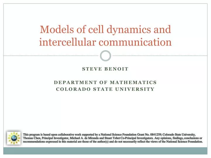
Models of cell dynamics and intercellular communication. Steve Benoit Department of Mathematics Colorado State University.

E N D
Models of cell dynamics andintercellular communication Steve Benoit Department of Mathematics Colorado State University This program is based upon collaborative work supported by a National Science Foundation Grant No. 0841259; Colorado State University, Thomas Chen, Principal Investigator, Michael A. de Miranda and Stuart Tobet Co-Principal Investigators. Any opinions, findings, conclusions or recommendations expressed in this material are those of the author(s) and do not necessarily reflect the views of the National Science Foundation.
Modeling Cell Dynamics & Communication Outline: • Context – CSU research program • Observing cell motion – phenomenology & analysis • Modeling cell behavior – top-down vs. bottom-up • New approaches at mesoscopic scale • Results & discussion
CSU Cell Communication Research Fluorescence Video Microscopy Analysis Optical Image Tissue Slice Preparation Tissue Sample Data Acquisition & Processing Electrochemical Biosensor Development Composite Image Biosensor Array Microchip Development Analysis Microchip Chemical Image Biomedical Sciences Chemistry Electrical & Computer Engineering Computer Science Mathematics
Cell Motion Analysis from Optical Images • Automated analysis tool: Z-planes } } } } Raw Image Merged Equalized Smoothed Stabilized Trajectories Analysis
Cell Motion Analysis from Optical Images • Automated analysis tool: Best-fit curves for model: α ≈ 1: diffusive behaviorα < 1: subdiffusive behavior α > 1: superdiffusive behavior α=.42 α=.93 α=1.25 Voronoi diagram and visualizations: Voronoi α K Speed Distance
Challenges of modeling cell behavior • Cell mechanics: Actin Microfilaments Intermediate Filaments Membrane & Adhesion Organelles Microtubules
Challenges of modeling cell behavior • Examples of cell motion & signaling: Neutrophil Macrophage Neuron T Cells Examples of cell mechanics: Actin motility Actin-InducedMovement MicrotubuleDynamics Adhesion
History: “Top-Down” Models • Continuum model of cell concentration (e.g. Keller & Segel -1971) • Random walk with bias (e.g. Alt – 1980) • Stochastic model(e.g. Tranquillo – 1988) • Hyperbolic continuum model(e.g. Hillen & Stevens - 2000)
History: “Bottom-Up” Models • Molecular dynamics models(~ 7x10¹⁵ atoms per cell) • Membrane models • Cytoskeleton models • Adhesion modulation models
Models for Mesoscopic Simulation • Plasma Membrane • Energy Functional Terms: • Axial displacement • Normal averaging • Radial Lennard-Jones • Each disc acts as sensor • ~500-1000 discs per cell • Disc Model • Triangulation • Simulation
Models for Mesoscopic Simulation • Organelles • Organelles are treated just as membranes • Different energy parameters • Interaction Terms: • Simple Lennard-Jones • Simulation
Models for Mesoscopic Simulation • Cytoskeleton • Collection of Lennard-Jones spheres • Interacts with other actin spheres and membrane discs • Interaction Terms: • Simple Lennard-Jones
Models for Mesoscopic Simulation • Cytoskeleton Dynamics & Signaling • Membrane discs are activated by signals • Actin spheres close to activated discs are activated / created • Activated spheres grow (polymerize) then divide • Global rate of sphere shrinkage (depolymerization) to restore monomer pool • Signals (Diffusive) • Simulation
Directions for Future Work • Parallelization and optimization • Simulations with 1000-5000 cells • Mitosos and apoptosis • Boundary/interior conditions (ventricle, radial glia)
Acknowledgements Colorado State University Tom Chen, Michael de Miranda CSU Biomedical Sciences Department and the Tobet Lab Stuart Tobet, Matt Stratton, KrystleFrahm, Brian Searcy CSU Department of MathematicsVakhtangPutkaradze University of Ljubljana & Jožef Stefan InstitutePrimožZiherl The author would like to thank the National Science Foundation for support under Grant No. 0841259.