Download
1 / 76
820 likes | 2.25k Views
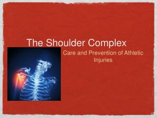
SHOULDER COMPLEX. Dr. Michael P. Gillespie. ARTICULATIONS OF THE SHOULDER. Four articulations of the shoulder exist involving the sternum, clavicle, ribs, scapula and humerus. The series of joints of the shoulder complex allow for extensive range of motion to the upper extremity.

E N D
SHOULDER COMPLEX Dr. Michael P. Gillespie
ARTICULATIONS OF THE SHOULDER • Four articulations of the shoulder exist involving the sternum, clavicle, ribs, scapula and humerus. • The series of joints of the shoulder complex allow for extensive range of motion to the upper extremity. • This extensive range of motion allows us to reach and manipulate objects. Dr. Michael P. Gillespie
JOINTS OF THE SHOULDER COMPLEX Dr. Michael P. Gillespie
MUSCLE INTERACTIONS • Muscles of the shoulder complex rarely act isolation, but rather work in “teams” to produce highly coordinated movements. • These movements are expressed over multiple joints. • The coordinated actions of multiple muscles allows for great versatility, control and range of motion. • Paralysis or weakness of any single muscle often disrupts the entire kinematic sequencing of the entire shoulder complex. Dr. Michael P. Gillespie
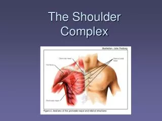
OSTEOLOGY • Sternum • Clavicle • Scapula • Proximal-to-Mid Humerus Dr. Michael P. Gillespie
OSTEOLOGIC FEATURES OF THE STERNUM • Manubrium • Pair of oval-shaped clavicular facets, which articulate with the clavicles. • Costal facets – attachment for first two ribs. • Jugular notch – superior aspect • Body • Xiphoid Process Dr. Michael P. Gillespie
STERNUM: ANTERIOR VIEW Dr. Michael P. Gillespie
OSTEOLOGIC FEATURES OF THE CLAVICLE • Shaft • Sternal end • Costal facet • Costal tuberosity • Acromial end • Acromial facet • Conoid tubercle • Trapezoid line Dr. Michael P. Gillespie
CLAVICLE: SUPERIOR AND INFERIOR SURFACES Dr. Michael P. Gillespie
OSTEOLOGIC FEATURES OF THE SCAPULA • Angles: Inferior, superior, and lateral • Medial or vertebral border • Lateral or axillary border • Superior border • Supraspinatous fossa • Infraspinatous fossa • Spine • Root of the spine • Acromion • Clavicular facet • Glenoid fossa • Supraglenoid and infraglenoid tubercles • Coracoid process • Subscapular fossa Dr. Michael P. Gillespie
SCAPULA: POSTERIOR & ANTERIOR VIEW Dr. Michael P. Gillespie
OSTEOLOGIC FEATURES OF THE PROXIMAL-TO-MID HUMERUS • Head of the humerus • Anatomic neck • Lesser tubercle and crest • Greater tubercle and crest • Upper, middle, and lower facets on the greater tubercle • Intertubercular (bicipital) groove • Deltoid tuberosity • Radial (spiral) groove Dr. Michael P. Gillespie
HUMERUS: ANTERIOR & SUPERIOR VIEWS Dr. Michael P. Gillespie
FOUR JOINTS WITHIN THE SHOULDER COMPLEX • Sternoclavicular • Acromioclavicular • Scapulothoracic • Glenohumeral Dr. Michael P. Gillespie
ARTHROLOGY OF THE SHOULDER COMPLEX • The most proximal articulation within the shoulder complex is the sternoclavicular joint. • The clavicle functions as a mechanical strut holding the scapula at a relatively fixed distance from the trunk. • The acromioclavicular joint attaches the scapula to the clavicle. • The anterior surface of the scapula rests against the posterior-lateral surface of the thorax, forming the scapulothoracic joint. This is not a true anatomic joint. It is an interface between bones. • The scapula serves as a base of operation for the glenohumeral joint. The glenohumeral joint is the most distal and mobile link of the complex. • “Shoulder movement” describes the combined motions at the glenohumeral and scapulothoracic joints. Dr. Michael P. Gillespie
PRIMARY MOVEMENTS OF THE SCAPULOTHORACIC JOINT • Elevation • Depression • Protraction • Retraction • Upward Rotation • Downward Rotation Dr. Michael P. Gillespie
ELEVATION & DEPRESSION OF THE SCAPULOTHORACIC JOINT Dr. Michael P. Gillespie
PROTRACTION & RETRACTION OF THE SCAPULOTHORACIC JOINT Dr. Michael P. Gillespie
UPWARD ROTATION & DOWNWARD ROTATION OF THE SCAPULOTHORACIC JOINT Dr. Michael P. Gillespie
STERNOCLAVICULAR JOINT: GENERAL FEATURES • The SC joint functions as the basilar joint of the entire upper extremity and links the appendicular skeleton to the axial skeleton. • The joint must be firmly attached, yet allow considerable range of movement. Dr. Michael P. Gillespie
TISSUES THAT STABILIZE THE STERNOCLAVICULAR JOINT • Anterior and posterior sternoclavicular ligaments • Interclavicular ligament • Costoclavicular ligament • Articular disc • Sternocleidomastoid, sternothyroid, sternohyoid, and subclavius muscles Dr. Michael P. Gillespie
KINEMATICS OF THE STERNOCLAVICULAR JOINT • Osteokinematics of the SC joint involve rotation in all three degrees of freedom. • Elevation & Depression • Protraction & Retraction • Axial (Longitudinal) Rotation of the Clavicle Dr. Michael P. Gillespie
OSTEOKINEMATICS OF THE STERNOCLAVICULAR JOINT Dr. Michael P. Gillespie
ACROMIOCLAVICULAR JOINT: GENERAL FEATURES • The AC joint is the articulation between the lateral end of the clavicle and the acromion of the scapula. • An articular disc is present in most AC joints. • The joint has an articular capsule and significant ligament support. Dr. Michael P. Gillespie
TISSUES THAT STABILIZE THE ACROMIOCLAVICULAR JOINT • Superior and inferior acromioclavicular joint ligaments • Costoclavicular ligament • Articular disc (when present) • Deltoid and upper trapezius muscles Dr. Michael P. Gillespie
KINEMATICS OF THE ACROMIOCLAVICULAR JOINT • The motions of the AC joint are described by the movement of the scapula relative to the lateral end of the clavicle. • Upward & Downward Rotation • Horizontal & Sagittal Plane “Rotational Adjustments” at the AC joint Dr. Michael P. Gillespie
OSTEOKINEMATICS OF THE ACROMIOCLAVICULAR JOINT Dr. Michael P. Gillespie
OSTEOKINEMATICS OF THE ACROMIOCLAVICULAR JOINT Dr. Michael P. Gillespie
ACROMIOCLAVICULAR JOINT DISLOCATION • The AC joint is inherently susceptible to dislocation due to the sloped nature of the articulation and the high probability of receiving large shearing forces. • Falling and striking the tip of the shoulder abruptly against the ground would produce such a shearing force. Dr. Michael P. Gillespie
SCAPULOTHORACIC JOINT • The scapulothroacic Joint is not a true joint, but rather a point of contact between the anterior surface of the scapula and the posterior-lateral wall of the thorax. • The two surfaces do not make direct contact. They are separated by muscles such as the subscapularis, serratus anterior, and erector spinae. • An audible click during scapular movements may indicate abnormal contact within the articulation. Dr. Michael P. Gillespie
KINEMATICS OF THE SCAPULOTHORACIC JOINT • The movements at the scapulothoracic joint are a result of cooperation between the SC and the AC joints. • Elevation & Depression • Protraction & Retraction • Upward & Downward Rotation Dr. Michael P. Gillespie
SCAPULOTHORACIC ELEVATION Dr. Michael P. Gillespie
SCAPULOTHORACIC PROTRACTION Dr. Michael P. Gillespie
SCAPULOTHORACIC UPWARD ROTATION Dr. Michael P. Gillespie
FUNCTIONAL IMPORTANCE OF UPWARD ROTATION • Many functional activities require us to raise the arm fully overhead. • The upward rotation of the scapula accounts for nearly 1/3 of the 180 degrees of shoulder abduction or flexion. • Functions • The upwardly rotated scapula projects the glenoid fossa upward and anterior-laterally, providing a structural base to maximize upward and lateral reach. • The upwardly rotated scapula preserves the optimal length-tension relationship of the abductor muscles of the glenohumeral joint (middle deltoid & supraspinatous). • The upwardly rotated scapula helps maintain the volume within the subacromial space. A reduced subacromial space can lead to painful and damaging impingement of the supraspinatus tendon and subacromial bursa). Dr. Michael P. Gillespie
GLENOHUMERAL JOINT: GENERAL FEATURES • The GH joint is the articulation formed between the large convex head of the humerus and the shallow concavity of the glenoid fossa. • It operates in conjunction with the moving scapula to produce an extensive range of motion of the shoulder. • In anatomic position, the articular surface of the glenoid fossa is directed anterior-laterally in the scapular plane. • In anatomic position, the humeral head is directed medially and superiorly, as well as posteriorly. • This orientation places the head of the humerus directly against the face of the glenoid fossa. Dr. Michael P. Gillespie
GLENOHUMERAL JOINT: ANTERIOR VIEW Dr. Michael P. Gillespie
“LOOSE FIT” OF THE GLENOHUMERAL JOINT & INSTABILITY • Several features of the glenohumeral joint contribute to a design that favors mobility at the expense of stability. • The articular surface of the glenoid fossa covers only about 1/3 of the articular surface of the humeral head. • The longitudinal diameter of the humeral head is about 1.9 times larger than the longitudinal diamter of the glenoid fossa. • The transverse diameter of the humeral head is about 2.3 times larger than the opposing transverse diameter of the glenoid fossa. • The surrounding muscles and ligaments maintain the mechanical integrity of the joint. • A condition of excessive laxity or “joint play” associated with large translations of the proximal humerus relative to the glenoid is often referred to as shoulder instability. • Subluxation – incomplete separation of articular surfaces often followed by spontaneous realignment • Dislocation – complete separation of articular surfaces without spontaneous realignment Dr. Michael P. Gillespie
“LOOSE FIT” IN GLENOHUMERAL JOINT Dr. Michael P. Gillespie
GLENOHUMERAL JOINT STABILITY • A combination of passive and active mechanisms achieve GH joint stability. • Active mechanisms • Forces produced by muscle • Embracing nature of the rotator cuff • Passive mechanisms • Forces other than activated muscle • 1. restraint provided by capsule, ligaments, glenoid labrum, and tendons • 2. mechanical support predicated on scapulothoracic posture • 3. negative intracapsular pressure Dr. Michael P. Gillespie
ROTATOR CUFF MUSCLES & LONG HEAD OF BICEPS BRACHII • The glenohumeral joint receives significant structural reinforcement from the four rotator cuff muscles. • The subscapularis is the thickest of these muscles and lies just anterior to the scapula. • The supraspinatus, infraspinatus, and teres minor lie superior and posterior to the capsule. • These four muscles form a cuff that protects and actively stabilizes the GH joint, especially during dynamic activities. • The belly of these muscles lies close to the joint. • The tendons of these muscle blend into the capsule. • The tendon of the long head of the biceps reinforces the rotator interval (between supraspinatus and subscapularis). Dr. Michael P. Gillespie
ROTATOR CUFF MUSCLE SUPPORT Dr. Michael P. Gillespie
TISSUES THAT REINFORCE OR DEEPEN THE GLENOHUMERAL JOINT • Joint capsule and associated capsular ligaments • Coracohumeral ligament • Rotator cuff muscles (subscapularis, supraspinatus, infraspinatus, and teres minor) • Long head of biceps brachii • Glenoid labrum Dr. Michael P. Gillespie
KINEMATICS OF THE GLENOHUMERAL JOINT • Movement occurs in all three degrees of freedom. • Abduction & Adduction • Flexion & Extension • Internal & External Rotation Dr. Michael P. Gillespie
OSTEOKINEMATICS OF THE GLENOHUMERAL JOINT Dr. Michael P. Gillespie
GLENOID LABRUM: VULNERABLE TO INJURY • The rim of the glenoid fossa is encircled by a fibrocartilage ring, or lip, known as the glenoid labrum. • It deepens the concavity of the fossa and increases the contact area with the humeral head to help stabilize the joint. • The superior part of the glenoid labrum is only loosely attached. • 50% of the fibers of the tendon of the long head of the biceps are direct extensions of the superior glenoid labrum. • Large or repetitive forces within the biceps tendon can detach the superior labrum (near its 12 o’clock position). Dr. Michael P. Gillespie
GLENOHUMERAL JOINT: ACTIVE ABDUCTION Dr. Michael P. Gillespie
GLENOHUMERAL JOINT: FLEXION Dr. Michael P. Gillespie
KINEMATIC RELATIONSHIPS OF THE GLENOHUMERAL JOINT Dr. Michael P. Gillespie
SCAPULOHUMERAL RHYTHM • “Scapulohumeral rhythm” describes the kinematic relationship between glenohumeral abduction and scapulothoracic upward rotation. • After about 30 degrees of abduction, the rhythm is remarkably constant. • For every 3 degrees of shoulder abduction, 2 degrees occur by GH joint abduction and 1 degree occurs by scapulothoracic upward rotation. • A full arc of 180 degrees of abduction is the result of a simultaneous 120 degrees of GH joint abduction and 60 degrees of scapulothoracic upward rotation. Dr. Michael P. Gillespie