Download
1 / 68
700 likes | 1.02k Views
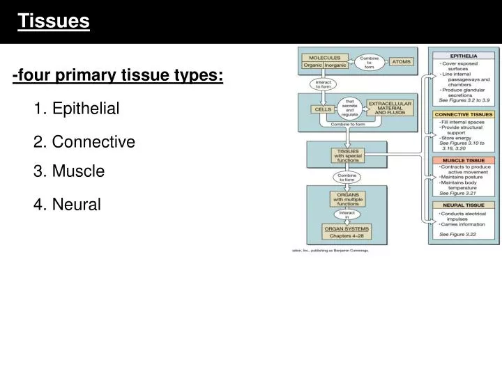
Tissues. -four primary tissue types:. 1. Epithelial. 2. Connective. 3. Muscle. 4. Neural. Epithelial Tissue. = lining epithelium & glands multiple functions of epithelial tissue: 1. protection - from dehydration, pathogens 2. synthesis 3. regulation - e.g. body temperature

E N D
Tissues -four primary tissue types: 1. Epithelial 2. Connective 3. Muscle 4. Neural
Epithelial Tissue • = lining epithelium & glands • multiple functions of epithelial tissue: • 1. protection - from dehydration, pathogens • 2. synthesis • 3. regulation - e.g. body temperature • 4. excretion - e.g. waste • 5. immune response • lining epithelium = line body surfaces and cavities • glandular epithelium = secretion
Three Major Functions of Epithelial Tissues 1. Physical protection: are found on exposed surfaces of the body -protection from abrasion, dehydration, and entrance by toxins 2. Control permeability: every substance that enters the body must cross an epithelial tissue first -permeability varies from location to location -contain pores, channels, transporters and other machinery required for selective permeability -function can be modified by stimuli e.g. hormones can increase ion transport e.g. stress can alter the physical structure and thus permeability 3. Sensitivity: innervated by sensory nerves -many epithelial tissues can detect differences in the environment (e.g. heat, pressure) -convey these changes to the nervous system -one type of specialized sensory epithelium = neuroepithelium -found in special sense organs (ear, eyes, tongue)
Epithelial characteristics • Cellularity • Polarity • Attachment • to each other • to connective tissue • Avascularity • Regeneration
Epithelial characteristics 1. Cellularity: composed almost entirely of cells held together by cell junctions -very little extracellular matrix 2. Polarity: possesses an exposed surface the faces the exterior of the body - apical face -also has an attached base which is anchored to other tissues - basal face -the organelles are not uniformly distributed
-the BM is comprised of two layers a. closest to epithelial cell = basal lamina (glycoproteins, laminin and actin) -acts as a barrier to transport b. furthest from the epithelial cell = reticular lamina -collagen IV bundles produced by the underlying connective tissue cells -provides strength Epithelial characteristics Epithelial characteristics 3. Attachment: attached to underlying tissues via the basement membrane -BM is produced by the basal surface of the cells & connective tissue - comprised of collagen type IV and laminin
1. Tight junctions: lipid portions of PMs are bound together by interlocking membrane proteins -very tight union - prevents passage of water and solutes between the two cells Epithelial characteristics Epithelial characteristics 3. Attachment: also form extensive connections between each other
3. Desmosomes: comprised of cellular adhesion proteins/CAMs and proteoglycans -also form plaques and contain cadherins -the plaque attaches to the intermediate filaments of the cytoskeleton (keratin) -several types known: belt, hemi, button -hemidesmosome: attaches the cell to the basement membrane of the tissue -link to a basement membrane protein = laminin
2. Gap junctions: two cells held together by proteins called connexons -connexons are channel proteins -materials can freely move between the two cells -passage of materials helps to coordinate the activities of the two cells e. g beating rhythm of cilia Epithelial characteristics Epithelial characteristics
Epithelial characteristics 4. Avascularity: do not contain blood vessels -must obtain nutrients via diffusion or absorption 5. Regeneration: damaged cells are replaced through differentiation of stem cells located deep within the tissue -rate of renewal depends on rate of cell death -stem cells = germinative cells -these cells are found closest to the basement membrane -migrate towards the surface and differentiate
Classification of Epithelia • catagorizing epithelial tissue types A. # of layers simple = 1 layer stratified = multiple **pseudo = 1 layer B. Cell shape columnar cuboidal squamous
Simple Epithelium Stratified Epithelium -relatively thin -cells have the same polarity - nuclei are generally aligned -very fragile - cannot provide mechanical/physical protection -line internal compartments -relatively permeable - absorptive surfaces, secretion, filtration -thicker due to multiple layers -found in areas subject to mechanical and chemical stress e.g. skin, mouth -tougher than simple epi.-organelles do not align
Pseudostratified Epithelium -appears to be stratified -yet the basal surface of every cell contacts basement membrane -apical surface of some cells may possess cilia -other cells secrete mucus -found lining absorptive organs e.g. respiratory epithelium
Squamous Epithelium -tile-like cells - cells are thin, flat and irregular in shape -cells interlock like tiles -simple squamous - most delicate tissue in the body -found in protected regions where absorption occurs -many types: mesothelium - lines ventral body cavity (i.e. abdomen) endothelium - lines heart and vessels
Squamous Epithelium -stratified squamous - where mechanical stresses are severe -cells on exposed surfaces contain keratin - an intermediate filament protein that reduces water loss and provides strength = keratinized epithelium -non-keratinized epithelium is tough but must be kept moist
Transitional Epithelium -permits stretching -located in walls of the bladder, renal pelvis and the ureters e.g. bladder wall - when empty the epi. looks as if it has several layers -actual number of layers can be seen upon distension
Cuboidal Epithelium -cells are cubes -nucleus is in the center of the cell -simple cuboidal: regions of secretion and absorption e.g. kidney tubules pancreas & salivary glands - buffers & enzymes thyroid follicles - thyroid hormones
Cuboidal Epithelium -stratified cuboidal: relatively rare -ducts of sweat glands and mammary glands
Columnar Epithelium -height is greater than their width -nuclei is close to the BM -simple columnar: provides some protection -also in areas of absorption and secretion
Simple columnar epithelium microvilli wandering lymphocytes -usually involved in secretion and absorption -located in the gallbladder, larger ducts of exocrine glands, gastric pits of stomach -basally located nuclei aligned with one another -frequently the apical face is modified with cellular extensions e.g. microvilli – intestinal lining = brush border -short-lived cells – replaced every 4 to 5 days -frequently found with Goblet cells (intestine and stomach)
Columnar Epithelium -stratified columnar: relatively rare -two to multiple layers -only outer layer contains truly columnar cells -protection role
Columnar Epithelium -pseudostratified columnar: only a single layer -every cell contacts the BM -nuclei are at varying levels - appearance of multiple layers -exposed apical surface typically bears cilia e.g. respiratory epithelium
Pseudostratified -these tissues are generally ciliated
Function: filtration, diffusion, osmosis secretion, absorption protection, secretion, absorption protection, secretion, movement of mucus protection Location: lungs,linings of blood vessels ovaries, kidneys, certain glands linings of uterus, stomach and intestines linings of respiratory passages and reproductive outer layer of skin, oral cavity, throat Type: Simple squamous Simple cuboidal Simple columnar Pseudostratified columnar Stratified squamous Description: single layer, flattened cells single layer, cube-shaped cells single layer, elongated cells single layer, elongated cells multiple layers, flattened cells
Glandular Epithelium • epithelial cells specialized to produce and secrete substances • gland = single epithelial cell OR multiple cells • two types of glands: 1) exocrine = secrete into ducts • e.g. sweat glands 2) endocrine = secrete directly into bloodstream e.g. thyroid, pituitary ** one gland is mixed - e.g. pancreas
exocrine gland structure: • Unicellular: single-celled glands • e.g. goblet cells • Multicellular glands – multiple cells grouped together • Can be classified based on: • 1. Mode of secretion – used by physiologists • Merocrine • Apocrine • Holocrine • 2. Consistency of secretion – used by histologists • Serous • Mucus • Mixed • 3. Structure – used by histologists • shape of the secretory portion • branching pattern of the duct • simplest multi-cellular gland is a secretory sheet • e.g. gastric epithelium
SIMPLE COLUMNAR with GOBLET CELLS -goblet cells = unicellular exocrine glands that secrete mucus
exocrine gland types • 1. serous - watery fluid that contains enzymes • e.g. saliva – parotid salivary gland
exocrine gland types • 2. mucous - glycoproteins called mucins that absorb water to form a • slippery mucus
exocrine gland types • 3. mixed - more than one type of gland cell • -produces different types of secretions - mucus and serous • e.g. submandibular or sublingual salivary gland
provides support • binds structures together • fills cavities • produces blood • protects organs • components: matrix + cells Connective Tissue -matrix: non-cellular support material -comprised of extracellular protein fibers – mainly collagens e.g. 1. collagen fibers (white) – type I 2. elastic fibers (yellow) 3. reticular fibers – collagen type III 4. fibronectin -plus a ground substance = water + hyaluronan (sugar), proteoglycans and glycoproteins -cells: secrete the matrix -some have become very specialized and make a very specialized matrix
Connective Tissues -cell types possibly found in connective tissues: A. fibroblasts: immature cell type found in basic connective tissues -secrete the extracellular matrix -secrete main component of matrix = collagens -also produce hyaluronan = glycosaminoglycan (sugar) that gives the ground substance a viscous quality -also produce the other components of the ground substance e.g. proteoglycans • adipocytes: main cellular component of adipose tissue • -more specialized type of fibroblast – fat storage • -fill with lipid upon maturation C. melanocytes: synthesize and secrete melanin -dark, brown pigment that absorb light
Connective Tissues D. Macrophages (Fixed): engulf damaged and dead cells by phagocytosis -immune cell -derived from monocytes E. free macrophages: wander rapidly through the connective tissue -called monocytes when circulating in blood F. mast cells: another immune cell -synthesize and secrete histamine - inflammation response -synthesize and secrete heparin - inflammation response G. lymphocytes - immune cells (T and B cells) -differentiate into plasma cells (type of B cells) - antibodies -differentiate into T cell subtypes - assist B cells
Connective tissue Matrix fibers: collagen, reticular and elastic 1. Collagen fibers: long, straight and unbranched fibers made of CN type I -very concentrated and dense in tendons and ligaments -long chains of collagen protein subunits forming a triple helix -these helices are wound together - “rope” or a bundle = fibril -fibrils are then stacked together = collagen fiber -triple helix – three subunits = 2 alpha 1 chains + 1 alpha 2 chain e.g. CNI - 2 chains of CNIa1 and one chain of CNIa2 -there are nineteen types of collagen in the body = 80-90% are CNI, CNII & CNIII
Connective tissue: fibers 2. Reticular: made of collagen type III -2 collagen III alpha1 + 1 collagen III alpha2 subunits = reticular fiber -reticular fibers interact in a different way – 3D network rather than bundles -thinner than collagen type I fibers - more flexible -abundant in the walls of hollow organs -form a supportive stroma (3D network) that supports the functional cells of these organs 3. Elastic: primarily made up of the proteins fibrillin & elastin -branching and wavy in appearance
Classification Embryonic: first to appear = mesenchyme -derived from mesoderm germ layer -cells are star-shaped -matrix - fine protein filaments -Mucus connective tissue - jelly-like, many regions of embryo
The way I organize Connective tissues • Loose – areolar CT, fat & reticular • Dense – dense (regular, irregular), elastic • Supportive – bone & cartilage • Fluid – blood & lymph
Type: Areolar Adipose Dense Cartilage Bone Blood Description: Cells in fluid-gel matrix Cells in fluid-gel matrix Cells in fluid-gel matrix Cells in solid-gel matrix Cells in solid matrix Cells in fluid matrix Function: Binds organs together, holds tissues, fluids Protects, insulates and stores Binds organs together Supports, protects, provides framework Supports, protects, provides framework Transports gases, defends against disease, clotting Types: 1. Areolar 2. Dense – regular and irregular 3. Adipose 4. Cartilage 5. Bone 6. Blood
Loose connective tissues: types 1. Loose Areolar tissue (Areolar tissue) -cells are mainly fibroblasts, spaced far apart -matrix: sparse collagen fibers, elastic fibers, mostly ground substance -cushions and can be distorted due to loose organization e.g. beneath the dermis
2. Adipose tissue = fat -cells = adipocytes (fat storing fibroblasts) -cushions joints and organs -stores energy -insulates liposuction: suction assisted lipoplasty -removal of SQ fat
3. Reticular: -thin collagen fibers in a 3D network (reticular fibers) -supports walls of certain organs e.g. liver, spleen
Dense connective tissues: types -most of the tissue is densely packed extracellular matrix fibers of collagen type I -often called collagenous tissue -type types: 1) dense regular - dense, elastic e.g. tendons, ligaments 2) dense irregular - interwoven meshwork or fibers -e.g. dermis of skin, perichondrium of joints and periosteum of bone
1. Dense: -few fibroblasts - multiple, closely packed collagen fibers - fine network of elastic fibers e.g. tendons, ligaments
Dense irregular tissue • Found in the deepest layers of the dermis = also called the reticular layer (don’t confuse it with reticular tissue)
2. Elastic: -yellow, elastic fibers in parallel or branching networks -walls of larger vessels, airways, hollow organs NOT ON YOUR PRACTICAL
Supportive Connective tissues: types -cartilage & bone 1. Cartilage: -cells = chondrocytes -matrix = collagen fibers embedded in a gel-like ground- substance -collagen type II -ground substance - water + proteoglycans -proteoglycans - protein + sugars e.g. chondroitin sulfate glucosamine -functions in support, attachment, protection -in developing child - model for future bone (endochondral bone) -avascular tissue - produces anti-angiogenic chemicals (inhibits growth of blood vessels) -therefore diffusion is the main mode of transport Proteoglycan
-3 types: 1) Hyaline - most common - “glass” - ends of bones, within joints (synovial, articular), - end of nose, supports respiratory passages