Download
1 / 26
260 likes | 403 Views
11.3 – The kidney. Human Health & Physiology. Kidney Structure & Function. Excretion: the process of removing metabolic waste from the cells, tissue fluid, and blood of living organisms The main organ of excretion in mammals is the kidney

E N D
11.3 – The kidney Human Health & Physiology
Kidney Structure & Function • Excretion: the process of removing metabolic waste from the cells, tissue fluid, and blood of living organisms • The main organ of excretion in mammals is the kidney • Osmoregulation: the control of water balance of the blood, tissue or cytoplasm of a living organism
Kidney Structure & Function • Functions: • Produces urine • Maintain water balance • Maintain blood pH • Maintain blood pressure • The functional unit of the kidney is the nephron • There are more than 1 million nephrons in a human kidney • On average 120mL/min of fluid passes through the kidney
Kidney Structure & Function • Roles of the nephron: • Ultrafiltration • Reabsorption • Secretion • For the kidney you should be able to label: • Cortex • Medulla • Pelvis • Ureter • Renal blood vessels
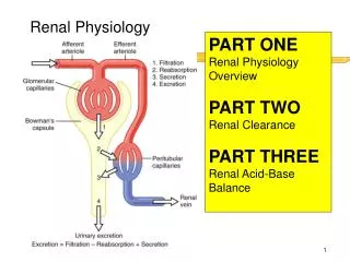
Nephron Structure & Function • Afferent arteriole: brings blood into the glomerulus from the renal artery • Efferent arteriole: takes blood out of the glomerulus into the surrounding capillary network and then into the renal vein • Glomerulus: a ball of capillaries that are fenestrated (have pores) and are surrounded by a basement membrane that filters what passes through into the filtrate
Nephron Structure & Function • Bowman’s capsule: a cup shaped structure at the end of the nephron that surrounds the glomerulus and collects the filtrate • Proximal convoluted tubule (PCT): lined with microvilli to increase surface area and has many mitochondria to provide ATP for active transport
Nephron Structure & Function • Loop of Henle: carries the filtrate from the PCT to the DCT • The loop of Henle descends into the medulla of the kidney • The concentration gradient of salt increases as you move down the medulla of the kidney
Nephron Structure & Function • Distal convoluted tubule (DCT): conducts urine from the loop of Henle to the collecting duct • It is the final place where blood pH and ions are balanced • Collecting duct: collects urine and carries it into the renal pelvis to the ureter • This is where the final water balance of the blood occurs
Formation of urine • Blood enters the glomerulus through the afferent arteriole under high pressure • This forces water, amino acids, small proteins, glucose, and ions into the Bowman’s capsule • The product is the filtrate that enters the nephron • Filtrate flows into the PCT • Glucose, amino acids, and ions are reabsorbed into the bloodstream through active transport • Small proteins are reabsorbed by pinocytosis
Formation of urine • Filtrate flows into the descending loop of Henle • The descending loop is permeable to water but not to salt • The loop of Henle is hypotonic (higher salt/urea concentration outside) to the medullary fluid • Water moves out of the nephron by osmosis • Therefore the filtrate becomes more concentrated and hypertonic
Formation of urine • The ascending loop of Henle is permeable to salt, but not to water • As the filtrate moves up the ascending loop, salt (Na+ and Cl–) moves out passively at first, then is actively pumped out at the top of the loop • Filtrate passes into the DCT where it is adjusted to balance blood pH (by secretion of H+) and ion composition • More water is also reabsorbed
Formation of urine • Filtrate moves into the collecting duct, where water may be further reabsorbed if needed • This is controlled by anti-diuretic hormone (ADH) • ADH is secreted by the pituitary gland and increases the permeability of the DCT and collecting duct to water
Formation of urine • If blood is low in water content, ADH is secreted and more water is reabsorbed from the collecting duct into the blood • Concentrated urine is formed and water is conserved • If blood is high in water content, ADH is not secreted and no more water is reabsorbed • Dilute urine is formed
Diabetes & Glucose in Urine • People with uncontrolled diabetes can have a large amount of glucose in their blood • Glucose enters the glomerular filtrate and is reabsorbed by active transport • There is a maximum rate at which reabsorption can occur • If there is too much glucose in the blood, reabsorption of all glucose from the glomerular filtrate cannot be achieved
References • Damon, A., McGonegal, R., Tosto, P., & Ward, W. (2007). Higher Level Biology. England: Pearson Education, Inc. • Raven, P.H., Johnson, G.B., Losos, J.B., Mason, K.A., & Singer, S.R. (2008). Biology. (8th ed.). New York: McGraw-Hill Companies, Inc.