Download
1 / 38
380 likes | 637 Views
Structural Bioinformatics. Proteins Secondary Structure Predictions. The first high resolution structure of a protein-myoglobin. Was solved in 1958 by Max Perutz John Kendrew of Cambridge University. (Won the 1962 and Nobel Prize in Chemistry ).

E N D
Structural Bioinformatics Proteins SecondaryStructure Predictions
The first high resolution structure of a protein-myoglobin Was solved in 1958 by Max Perutz John Kendrew of Cambridge University. (Won the 1962 and Nobel Prize in Chemistry) In 12.12.2013 there were 89,110 protein structures in the protein structure database. Great increase but still a magnitude lower then the total number of protein sequence databases (close to 1,000,000)
What can we do to bridge the gap?? MERFGYTRAANCEAP…. Predicting the three dimensional structure from sequence of a protein is very hard (some times impossible) However we can predict with relative high precision the secondary structure
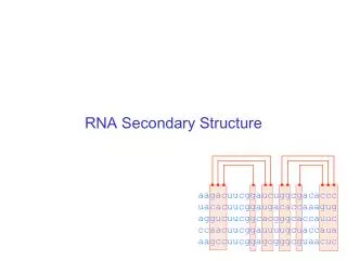
What do we mean bySecondary Structure ? Secondary structure are the building blocks of the protein structure: =
What do we mean bySecondary Structure ? Secondary structure is usually divided into three categories: Anything else – turn/loop Alpha helix Beta strand (sheet)
The different secondary structures are combined together to form theTertiary Structure of the Proteins
Secondary Tertiary ? ? RBP ? Globin
Secondary Structure Prediction • Given a primary sequence ADSGHYRFASGFTYKKMNCTEAA what secondary structure will it adopt (alpha helix, beta strand or random coil) ?
Secondary Structure Prediction Methods • Statistical methods • Based on amino acid frequencies • HMM (Hidden Markov Model) • Machine learning methods • SVM , Neural networks
Statistical Methods for SS prediction Chou and Fasman (1974) Name P(a) P(b) P(turn) Alanine 142 83 66 Arginine 98 93 95 Aspartic Acid 101 54 146 Asparagine 67 89 156 Cysteine 70 119 119 Glutamic Acid 151 037 74 Glutamine 111 110 98 Glycine 57 75 156 Histidine 100 87 95 Isoleucine 108 160 47 Leucine 121 130 59 Lysine 114 74 101 Methionine 145 105 60 Phenylalanine 113 138 60 Proline 57 55 152 Serine 77 75 143 Threonine 83 119 96 Tryptophan 108 137 96 Tyrosine 69 147 114 Valine 106 170 50 The propensity of an amino acid to be part of a certain secondary structure (e.g. – Proline has a low propensity of being in an alpha helix or beta sheet breaker) Success rate of 50%
Secondary Structure Method Improvements ‘Sliding window’ approach • Most alpha helices are ~12 residues longMost beta strands are ~6 residues long • Look at all windows of size 6/12 • Calculate a score for each window. If >threshold predict this is an alpha helix/beta sheet TGTAGPQLKCHIQWMLPLKK
Improvements since 1980’s • Adding information from conservation in MSA • Smarter algorithms (e.g. Machine learning, HMM).
HMM (Hidden Markov Model) approach for predicting Secondary Structure • HMM enables us to calculate the probability of assigning a sequence to a secondary structure TGTAGPOLKCHIQWML HHHHHHHLLLLBBBBB p = ?
Beginning with an α-helix The probability of observing Alanine as part of a β-sheet α-helix followed by α-helix The probability of observing a residue which belongs to an α-helix followed by a residue belonging to a turn = 0.15 Table built according to large database of known secondary structures
Example What is the probability that the sequence TGQ will be in a helical structure?? TGQ HHH p = 0.45 x 0.041 x 0.8 x 0.028 x 0.8x 0.0635 = 0.0020995 Success of HMM based methods-> 75%-80%
Mad Cow DiseasePrPc to PrPsc PRPc PRPsc
How do the protein structure relate to the primary protein sequence??
SEQUENCE -Early experiments have shown that the sequence of the protein is sufficient to determine its structure (Anfisen) - Protein structure is more conserved than protein sequence and more closely related to function.
How (CAN) Different Amino Acid Sequence Determine Similar Protein Structure ?? Lesk and Chothia 1980
Different sequences can result in similar structures 1ecd 2hhd
We can learn about the important features which determine structure and function by comparing the sequences and structures ?
Where are the gaps?? The gaps in the pairwise alignment are mapped to the loop regions
retinol-binding protein odorant-binding protein apolipoprotein D How are remote homologs related in terms of their structure? RBD b-lactoglobulin
PSI-BLAST alignment of RBP and b-lactoglobulin: iteration 3 Score = 159 bits (404), Expect = 1e-38 Identities = 41/170 (24%), Positives = 69/170 (40%), Gaps = 19/170 (11%) Query: 3 WVWALLLLAAWAAAERD--------CRVSSFRVKENFDKARFSGTWYAMAKKDPEGLFLQ 54 V L+ LA A + S V+ENFD ++ G WY + K Sbjct: 1 MVTMLMFLATLAGLFTTAKGQNFHLGKCPSPPVQENFDVKKYLGRWYEIEKIPASFE-KG 59 Query: 55 DNIVAEFSVDETGQMSATAKGRVRLLNNWDVCADMVGTFTDTEDPAKFKMKYWGVASFLQ 114 + I A +S+ E G + K V + ++ +PAK +++++ + Sbjct: 60 NCIQANYSLMENGNIEVLNKELSPDGTMNQVKGE--AKQSNVSEPAKLEVQFFPL----- 112 Query: 115 KGNDDHWIVDTDYDTYAVQYSCRLLNLDGTCADSYSFVFSRDPNGLPPEA 164 +WI+ TDY+ YA+ YSC + ++ R+P LPPE Sbjct: 113 MPPAPYWILATDYENYALVYSCTTFFWL--FHVDFFWILGRNPY-LPPET 159
The Retinol Binding Protein b-lactoglobulin
Taken together MERFGYTRAANCEAP…. FUNCTION
Pfam Database that contains a large collection of multiple sequence alignments of protein families (common structures) Very useful for function prediction. http://pfam.sanger.ac.uk/
The zinc-finger family (domain) Known family of Transcription Factors Protein sequence ZINC FINGER DOMAIN
Pfam Based on Profile hidden Markov Models (HMMs) which represents the protein family HMM in comparison to PSSM is a model which considers dependencies between the different columns in the matrix (different residues) and is thus much more powerful!!!! http://pfam.sanger.ac.uk/
Profile HMM (Hidden Markov Model)can accurately represent a MSA D19 D16 D17 D18 100% 16 17 18 19 delete D R T R D R T S S - - S S P T R D R T R D P T S D - - S D - - S D - - S D - - R 100% 50% M16 M17 M18 M19 100% 100% 50% Match D 0.8 S 0.2 P 0.4 R 0.6 R 0.4 S 0.6 T 1.0 I16 I17 I18 I19 insert X X X X
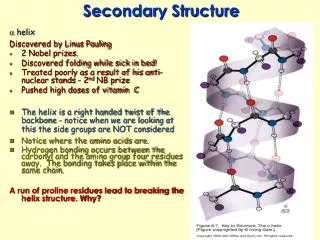
Alpha Helix: Pauling (1951) • A consecutive stretch of 5-40 amino acids (average 10). • A right-handed spiral conformation. • 3.6 amino acids per turn. • Stabilized by Hydrogen bonds 3.6 residues 5.6 Å
Beta Strand: Pauling and Corey (1951) β -strand > An extended polypeptide chains is called β –strand (consists of 5-10 amino acids > The chains are connected together by Hydrogen bonds to form b-sheet β -sheet
Loops • Connect the secondary structure elements (alpha helix and beta strands). • Have various length and shapes.