Download
1 / 42
520 likes | 1.45k Views
Neonatal & Pediatric Blood Gases. JOHN J ANCY, MA, RRT SENIOR CLINICAL CONSULTANT. Neonatal & Pediatric Blood Gases. Understanding neonatal & pediatric BGs Fetal/placental circulation Changes at birth Common neonatal cardiopulmonary misfires Cord, capillary, neonatal BGs Pediatric BGs.

E N D
Neonatal & Pediatric Blood Gases JOHN J ANCY, MA, RRT SENIOR CLINICAL CONSULTANT
Neonatal & Pediatric Blood Gases Understanding neonatal & pediatric BGs • Fetal/placental circulation • Changes at birth • Common neonatal cardiopulmonary misfires • Cord, capillary, neonatal BGs • Pediatric BGs
Fetal Circulation • Fetal nutrition, gas exchange, acid-base, electrolyte, fluid balance & waste removal rely on placental circulation • Fetal lungs not involved in gas exchange • Fetal circulation is markedly different than extra-uterine circulation • In-utero fetus exists in hypoxic state • At birth, breathing and circulatory changes must occur rapidly, if not-BIG problems in a little baby
Placental Structure • Gases , fetal waste and nutrients exchange across placenta • Maternal & fetal circulation physically separate • Placental BGs (fetal side) pH 7.30-7.40 pCO2 34-48 pO2 22-34 • Maternal CO↑30% • Maternal CV problems can occur late in pregnancy & Effect baby’s health
Fetal Circulation • Placenta • Umbilical vein oxygenated blood • Ductusvenosus carries blood to R Atrium • Foramen Ovale shunts blood from R Atrium to L Atrium • Ductusarteriosus bypasses lungs by connecting pulmonary artery to aorta • Umbilical arteries deoxygenated blood Foramen Ovale
Physiology of Fetal Circulation • Fetal Blood flow • Fetal lungs are non functioning-high PVR = circulatory bypass • Umbilical Vein carries oxygenated placental blood to fetus via Inferior Vena Cava • Most blood passes through D. Venosus to R Atrium bypassing liver • Blood entering R Atrium passes through Foramen Ovaleto L Atrium to L Ventricle sending oxygenated blood to head & upper limbs via Aortic Arch & bypasses lungs (Rt to Lt Shunt) • Deoxygenated blood returns to R Atrium via Superior Vena Cava to right ventricle to pumonary artery with majority of blood shunting through D. Arteriosusto Descending Aorta. • From Aorta to Umbilical Arteries deoxygenated, waste containing to placenta
Role of Fetal Hemoglobin-HbF • Fetal Hemoglobin has greater O2 affinity than HbA, left shift maximizes O2 transfer from maternal blood to fetal blood • HbA 2ɑ2Ƅ • HbF 2ɑ2ʏ • Fetal HbA production starts last trimester • HbF persists up to 8 months • Preemies have more HbF • Can compromise O2 delivery in preemies
Circulation Changes at Birth • Circulatory changes start with the first breath and rapidly alters with subsequent breaths • As lungs expand, PVR drops opening Right Heart circulation. Ventilation and pulmonary circulation are simultaneous, codependent. • Anything that compromises ventilation interferes with changes in circulation • Hypoxia, asphyxia, aspiration, atelectasis, etc can partially reverse circulatory changes and put baby in harm’s way • Cord BGs used in assessment of risk
Cord Blood Gases • Immediately at birth, cord is double clamped, cord arterial (fetal status) cord venous (placental status) • Indication: APGAR < 7 at 5 minutes • Reference ranges vary by study and gestation • Critical pH value is most often used
Cord Blood Reference Ranges Umbilical Arterial Critical Value pH < 7.20
Cord Blood Sample Handling Quality and accuracy of result requires correct handling • Should be drawn within 5 minutes of clamping • Labeled as Cord arterial or Cord venous • Use heparinized syringe (dry Li-Hep) • Mix immediately & prior to analysis • Must be air bubble free • Non-iced if analyzed within 30 minutes • Iced 30 to 60 minutes
Decrease in PVR at birth • During fetal circulation, lungs receive 5-10% of circulation, • In normal birth transition, 10X pulmonary blood flow increase • PVR decrease at birth • Lung distension • Decreased pCO2 and increased pO2 • Increase in lung NO levels near end of gestation
Circulation Changes at Birth • As fetus moves through birth canal, thorax is squeezed. Helps express amniotic fluid from lungs and spreads surfactant. • Premature infants have lower volume of less active surfactant and are more prone to IRDS and atelectasis • Surfactant reduces surface tension • C-Section babies have more residual amniotic fluid in lungs and less efficient surfactant distribution • Lungs mature later in development (more so in males) • These and many other conditions can inhibit birth circulatory changes secondary to elevated PVR
Circulation Changes at Birth • Placental circulation cut, ending cord blood flow • First breaths open lungs, lung expansion, oxygenation, pulmonary capillary dilation decrease PVR • Left atrial pressure increases dramatically, now higher than right atrial pressure • Functionally closes foramen ovale = reversing Rt–Lt shunt • Oxygenated blood in aorta constricts smooth muscle of ductusarteriosus = functional closure
Some things that can go wrong • Premature birth or gestational diabetes • Immature lungs = surfactant/gas exchange problems • Relative hypoxia = increased PVR = R-L shunt associated with incomplete conversion or reversal to fetal circulation • Even tracheal suctioning can cause increases in R-L shunt in some infants • Eclampsia- high BP, proteinuria, seizures • Occurs in 7% of pregnancies • Cardiopulmonary Anomalies • 1/1000 babies have a serious cardiac anomaly
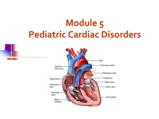
Most Common Cardiac Anomalies • Ventricular Septal Defect (VSD) 28% • Atrial Septal Defect (ASD) 11% • Pulmonary Stenosis 9% • Patent Ductus Arteriosus 9% (often occurs with other anomalies) • Aortic Stenosis 7% • Aortic Coarctation 6% • Surgical Intervention usually required
Some Neonatal Survival Threats • TTN • PPHN • IRDS • Congential diaphragmatic hernia • Meconium Aspiration Syndrome • Pneumonia • Pneumothorax • Cardiopulmonary anomalies • Respiratory Distress Syndrome • Sepsis Typical BGs = Respiratory Acidemia, mixed acidemia & hypoxemia
TTN-Transient Tachypnea of the Newborn • Delayed lung fluid clearance • Occurs in 0.3 to 0.5% of newborns • C-Section, maternal asthma, smoking & male gender • Self-limiting in 24 to 72 hours • Increased O2 requirement • ABGs reflect normal to mildly decreased pCO2 and hypoxemia
PPHNPersistent Pulmonary Hypertension of the Newborn • Failure of normal circulatory transition • Caused by Pulmonary Hypertension • R to L Shunt from patent Ductus Arteriosus and Foramen Ovale • Result = fetal circulation not completely altered • Refractory hypoxemia, respiratory distress and acidemia • Upper torso continues to receive better oxygenation • Most in near term infants, less in preemies • Severe PPHN 1-3 in 500 newborns • PPHN occurs in 10% of infants with respiratory failure
Causes of PPHN • Acute pulmonary hypertension • Alveolar hypoxemia (meconium, IRDS, pneumonia) • Hypoventilation (from asphyxia or neurologic conditions) • Hypothermia • Hypoglycemia
Types of PPHN • Abnormalities of lung parenchyma • Meconium aspiration • Pneumonia • Infant respiratory distress syndrome • Hypoplastic pulmonary vasculature • Diaphragmatic hernia • Idiopathic (10-20%)
BGs in PPHN • Extrapulmonary R-L shunting • Respiratory acidemia to mixed acidemia • Refractory hypoxemia • Elevated lactate • Right arm(preductal) pO2 & SpO2 higher than lower limbs (postductal) values - left arm varies • Hypoglycemia makes gas exchange worse • Hypocalcemia inhibits NO production = increased PVR • Monitor MetHb when iNO is in use • Cardiac anomalies can appear as PPHN
Treatment of PPHN (& others) • Monitor oxygenation, BP and perfusion • Continuous pulse oximetry preductal & postductal • Inotropic drugs • Surfactants (MAS, Sepsis, IRDS) • Mechanical ventilation (maintains/improves FRC) • Peak Pressure <30cmH2O & MAP <15cm H2O • 9 rib ventilation (X-Ray) • BG strategy-pO2 around 50mmHg is OK, use PEEP keep FiO2 as low as possible • BG strategy-correct respiratory acidosis (pCO2 up to 60) • Metabolic acidosis Bicarb if pCO2 OK THAM if not
Treatment of severe PPHN • High frequency ventilation • iNO – inhaled nitrous oxide (more on next slides) • Oxygenation index > 25 OI = (FiO2 x MAP cmH2O)/pO2 FiO2 0.60 MAP 22 pO2 50 OI = 26.4 = (60 x 22)/50 • iNO has reduced need for ECMO by 40% • ECMO • OI >40 e.g. FiO2 65, MAP 30, pO2 45: OI = 43 • Weight > 2000g • No major intracranial hemorrhage
Therapeutic use of iNO • Inhaled Nitric Oxide is primarily used for tx of PPHN (persistent pulmonary hypetension of the newborn) FDA approved • PPHN characterized by pulmonary hypertension and severe hypoxia secondary to right to left shunting through ductus arteriosus and/or foramen ovale • iNO acts as selective pulmonary vasodilator
Actions of iNO • iNO diffuses across alveolar membrane • Relaxes vascular smooth muscle • Reduces pulmonary vascular resistance • Reduces right to left shunting • Improvemes in oxygenation • iNO must be administered continuously due to transitory effect • iNO forms MetHb (most centers consider 5 to 7% MetHb as marker for iNO toxicity)
iNO use Guidelines • Lungs must be recruited and expanded • Consider HFV if FRC is low and recruitment difficult • iNO starts at 20 ppm, wean in 5 ppm increments, • Discontinue at FiO2 40-60 & iNO @ 1 ppm • Do not discontinue at higher doses due to rebound potential • The current detection level for iNO toxicity is 5 to 7%
IRDS- Infant Respiratory Distress Syndrome Surfactant deficiency = IRDS • Complex lipoprotein, produced by Type 2 alveolar cells • Surfactant production begins late in gestation • Reduces surface tension = prevents atelectasis • Occurs with premature birth, gestational diabetes, more often in male infants • Surfactant production/distribution is impaired by hypoxia, acidosis, hypotension & hypothermia
IRDS • 20000 to 30000 cases in US or 1% of births • 26-28 weeks gestation- 50% develop IRDS • 30-31 weeks gestation- 33% develop IRDS • More common with lower birth weights • White male infants • Diabetic mothers • Infants born by means of cesarean delivery • Second-born twins • Infants with a family history of respiratory distress syndrome
IRDS Deficient surfactant is the cause: • Atelectasis • V/Q mismatch • Hypoventilation & impaired gas exchange • Endothelial & epithelial disruption leads to proteinaceous leakage and pinkish hyaline membrane formation • Patent Ductus Arteriosus can complicate treatment course
IRDS treatment • Antenatal steroid administration • Surfactant administration • Has reduced mortality by 50% • Nasal CPAP and O2 support • High flow heated O2 therapy • Mechanical ventilation • Protective strategies (PP < 30 MAP < 15) • Lowest effective FiO2 (keep pO2 50-70) • Consider permissive hypercapnia • High frequency ventilation • iNO in selected cases (pulmonary hypertension)
IRDS BGs • Respiratory and mixed acidemia (frequent) • Moderate to severe hypoxemia & refractory • Elevated lactate • Typical BG: pH 7.23 pCO2 49 pO2 48 HCO3 19.8 BE -6.6 lactate 5.8 FiO2 60 PEEP 7 PP 24 MAP 14
Pediatric Respiratory Disease • Infections Typical BG, Repiratory Alkalemia, normoxia to mild to moderate hypoxemia • Pneumonia • Epiglottis • Bacterial Tracheitis • Croup • Asthma BGs- Respiratory Alkalemia with mild hypoxemia to Respiratory or Mixed Acidemia with moderate to severe hypoxemia. Lactate >4.0 mmol and/or Hyperkalemia may be present. Continuous neb Albuterol can reduce K, but can cause transient Hypokalemia
Pediatric Respiratory Disease • Bronchopulmonary Dysplasia BGs- Neonatal vs Pediatric Respiratory Acidosis to Chronic Respiratory Acidosis Varying degrees of Hypoxemia • Cystic Fibrosis BGs- Hypoxemia, none to mild to moderate CO2 retention Respiratory Acidosis and hypoxemia during exacerbations/pneumonia
Normal Range Adapted from: Deorari, AIIMS, 2008
ABGs Vary with Age Adapted from: Deorari, AIIMS, 2008
ABGs Vary with Gestational Age Adapted from: Deorari, AIIMS, 2008
Capillary BGs • Capillary samples if obtained from free flowing and pre-warmed site (arterialized) will closely reflect Arterial values for pH and pCO2. • pcO2 correlates well with paO2 if arterial is 60mmHg or less • Draw site should be warmed to 42C for 5 minutes, increases flow 7X. • Discard first drop, mix well & cap tube. • Remember heel cap samples might yield different results than arterial if PFC present
Arterial Sampling Precautions • Use dry Lithium heparin syringes only, 1.0 cc for pediatrics if ABGs with electrolytes • For BGs/electrolytes, use low concentration or balanced heparin. Fill syringes to 50% or more, otherwise Na and iCa will be falsely lowered • Remove entrapped air immediately • Mix immediately in two planes for >30 sec • Remix immediately prior to analysis • Icing not required if analyzed <30 minutes • Ice 30 minutes to 1 hour
Umbilical Arterial Catheter • Can be placed up to 48 Hrs post birth • Remove 2-3x line deadspace volume for waste draw • Use heparinized syringe only • For BGs/electrolytes, use low concentration or balanced heparin. Fill syringes to 50% or more, otherwise Na and iCa will be falsely lowered • Remove entrapped air immediately • Mix immediately in two planes for >30 sec • Remix immediately prior to analysis
Summary at Last • Understanding normal fetal circulation and birth transition helps in understanding what can go wrong and treatment course • Normal values vary with age and gestational age at birth • White males are more susceptible to perinatal respiratory complications • Proper sample handling is critical for reporting accurate values and reducing potential for treatment error