Download
1 / 43
430 likes | 442 Views
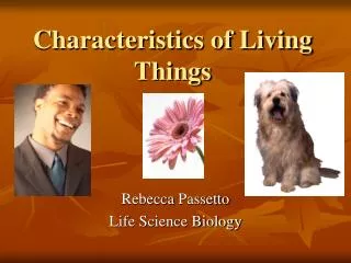
Learn about the concepts that define living things and explore the structure and functions of cells. Discover how microscopes revolutionized biology and the different parts of a cell that can be seen under a microscope.

E N D
DO NOW: Write down 5 things (concepts) that identifies if something is living or non living. AIM: What is a cell and what are its parts?
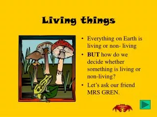
1) What is life? • In Biology, life is defined not by what it IS, but by what it DOES. • We call something that is alive an organism. • There are 8 life functions that living things do that separate them from non-living things.
2) What are the 8 life functions? • Nutrition- the ability to obtain and digest food • Respiration- the ability to convert food (organic molecules) into energy. • Synthesis- the ability to make molecules that the organism cannot get from the environment. • Growth- the ability to increase in size • Transport- the ability to move materials into, out of and throughout an organism.
What are the 8 life functions? (cont.) • Excretion- the ability to remove waste products made by cells. • Regulation- the ability to maintain homeostasis. - Homeostasis- a stable internal environment in a changing external environment. • Reproduction- the ability of an organism to produce more of its own kind Not needed for the individual organism to survive, but needed for the species to survive.
Because the cell can perform all of the 8 life processes, it is considered the smallest unit of life. 3) What is the smallest unit of life?
4) Do you need cells in order to be alive? • All organism, big or small, must contain at least one cell. • With out these cells, we could not perform any of the 8 life functions.
Don’t Copy!! When was the cell discovered? Until the invention of the microscope in 1590, no one ever seen or knew about cells. It was until 1663, when Robert Hooke looked at a thin slice of cork and coined the phrase “cell” which means small rooms.
5) The Cell Theory • Every organism is made up of one or more cells. • The cell is the basic unit of structure and function in all living things. • All cells come from pre-existing (old) cells. There are 3 rules in the cell theory:
6) Are there different groups of cells? • Cells can be grouped into two categories: - Prokaryote: these are cells that do not have a nucleus. Example: Bacteria - Eukaryote: these cells have a distinct nucleus.
Prokaryote Cell Eukaryote Cell Nucleus No Nucleus
7) What are the types of Eukaryote cells? • Eukaryotic cells fall into two categories: • a) Plant Cells b) Animal Cells
8) What are the parts of the cell that you can see under a microscope? • The cell is made up of many different parts. Each part does a specific job. The parts work together allowing the cell to survive. These parts are called organelles. • Organelles are specialized parts of the cell that perform a specific job.
DO NOW: Today’s televisions are said to be better than televisions of the past. Write down what you think “high definition” means. Day 12 AIM: The Cell - What you can see through a microscope? http://www.youtube.com/watch?v=Q3B3OnTVvmg
How did the microscope change biology? • Before the invention of the microscope, Scientists could not see cells, bacteria and protozoans (unicellular organisms)
The Light Microscope • The earliest microscope was the light microscope. • It used light from the sun, candles and later the light bulb. • Light would move from the bottom of the microscope, though the specimen (the thing you are looking at) and then through a series of lens. • The light microscope magnified the specimen, making it appear larger.
What could you see with a light microscope? • The best light microscopes can magnify a specimen up to 2000 times its size. • A skilled scientist, using various stains and techniques, can see many organelles inside a cell.
What could you see with a light microscope?Cell Membrane • The cell membrane: FUNCTION: • surrounds and protects the cell • controls what goes in and out (the bouncer) STRUCTURE: - is made of a double-lipid layer
What could you see with a light microscope?Nucleus • The nucleus: FUNCTION: • Controls all of the activities of the cell (the control center) STRUCTURE: - enclosed by its own membrane and contains the cell’s DNA
What could you see with a light microscope? Cytoplasm • The cytoplasm: FUNCTION: • is where the cell’s chemical reactions take place • It’s the transport system of the cell STRUCTURE: - as a fluid-like material consistently mainly of water
What could you see with a light microscope? Cell Wall • The cell wall: FUNCTION: • Gives the cell strength and rigidity • Does not interfere with the passage of materials into/out of the cell STRUCTURE: - Is made of cellulose NONE
What is an electron microscope? • In 1928, two German scientists invented a microscope that can magnify an image 2 millions times. This microscope was called an electron microscope. • Unlike a light microscope that use light to filter through a specimen. An electron microscope bombards the specimen with electrons.
What can you see with the electron microscope? • Because the electron microscope can magnify the specimen 1000 of times better than a light microscope with better resolution, we can see inside the cell with great detail. Do Not Copy Animal through light microscope Animal through electron microscope
Organelles you can see with an Electron Microscope. Except for the nucleus, chloroplast, and the cell membrane, most organelles are too small to see with a light microscope. The Electron Microscopecan focus energy so tightly that it an focus on very tiny things, like organelles. Do Not Copy
Mitochondria • Mitochondria are organelles the convert glucose into energy (ATP) • Mitochondria are found in the cytoplasm of the cell. • Cells that are very active, like muscle cells, have lots of mitochondria because they need lots of energy. • They are found in both animal and plant cells.
Mitochondria Draw
Ribosomes • Ribosomes are small circles of membrane located in the cytoplasm on the endoplasmic reticulum (ER). • Ribosomes are the site where amino acids are bonded together to form protein chains.
Ribosomes Draw
Endoplasmic Reticulum • The Endoplasmic Reticulum (ER) is a series of channels or channels that help in the transport of large molecules, such as proteins, in and around the cell. • There are 2 types of ER • Rough ER which has ribosomes attach to it. • Smooth ER which has no ribosomes.
Vacuole • The vacuole is a chamber in the cell. • In plants the vacuole is large and toward the center of the cell. • In animals, the vacuoles are small or mot there at all. • It holds extra food or water • It plants the vacuole helps give the plant support.
Chloroplast • Chloroplast are only found in plant cells. • This is the site of photosynthesis. • They change light energy into stored chemical energy (glucose). • They are found in the cytoplasm.
Place a check (√) in the box if the cell has that structure.
Place a check (√) in the box if the cell has that structure.
Anacharis cell (plant cell) cytoplasm chloroplasts Cell wall
Human cheek cell (animal cell) cytoplasm nucleus Cell membrane