Download
1 / 17
250 likes | 698 Views
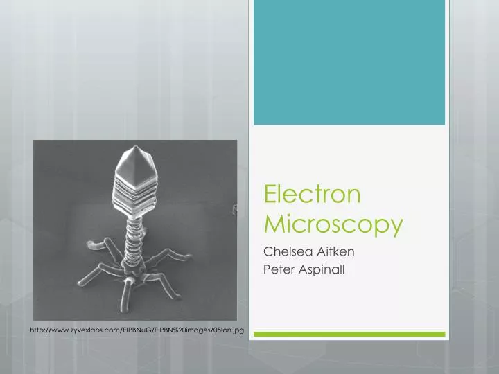
Electron Microscopy. Chelsea Aitken Peter Aspinall. http://www.zyvexlabs.com/EIPBNuG/EIPBN%20images/05Ion.jpg. Intro to Electron Microscopy. Similar to optical microscopy except with electrons rather than photons Used to image samples with a resolution of 10 Å

E N D
Electron Microscopy Chelsea Aitken Peter Aspinall http://www.zyvexlabs.com/EIPBNuG/EIPBN%20images/05Ion.jpg
Intro to Electron Microscopy • Similar to optical microscopy except with electrons rather than photons • Used to image samples with a resolution of 10 Å • Can image many different structural geometries • Mostly limited by radiation damage from the electron beam
Electron Properties • Since electrons exhibit wave and particle behavior, the de Broglie relationship applies: • Since the electron is charged, when introduced to an electric potential difference, it accelerates to its equilibrium momentum: • So particle momentum is only dependent on the electric potential difference
Electromagnetic Lenses • Used to focus the electron beam • We can relate wavelength to accelerating voltage: • Electron wavelengths are 5 times smaller photons • Maximum resolution (d) of a lens is related to the aperture angle and the wavelength by: • However due to aberration in the lens, the resolution is also limited by: • Where Cs is the spherical aberration coefficient
Signal vs. Noise • Largest issue is radiation damage to the specimen • Image is generated from elastic scattering while noise is generated from the inelastic scattering • Inelastic scattering deposits energy on the sample which damages the sample (occurs 3-4 times more often than elastic scattering events) • Signal-to-noise ratio is described by the Rose model: • Where C is the contrast () • Ratios between 5-7 are required to identify features with good enough confidence • Can either use an energy filter (remove certain energies) or higher accelerating voltage
Effect of the Microscope on Electron • When a wave is passed through a samples, it interacts and is released as a phase shift of the wave in. This is represented by: • This is then adjusted to account for electron absorbance: • The microscope then observes the phase shift due to this change which can be represented by the Fourier Transform: • Where P(u,v) is the transfer function of the microscope
Electron Generation • Thermionic Electron Gun • Heated filament produces electrons • Typically made of Tungsten or Lanthanum hexaboride • Electrons drawn towards an anode • An aperture in the anode creates a beam • Field Emission Gun • A very strong electric field is used to extract electrons from a metal filament • Filament typically a single tungsten crystal • Requires a vacuum • Similar anode setup
Microscope Setup • Transmission Electron Microscope • Phase contrast Image is formed by the interference between electrons that passed through the sample and ones that did not • Scanning Electron Microscope • Electron beam is scanned across the sample • The reemitted electrons are measured in order to form the image
Focusing • When the image is in focus, there is very low contrast due to the electron loss around the objective • By imaging underfocus or overfocus, a phase shift and amplitude contrast are created • This creates a dark image with a white ring around or a white image with a dark ring (respectively
Negative Staining • Biological samples are often imaged using negative staining • The elements of biological molecules do not interact strongly with the electron beam • Instead they are seated in a material that does and then the negative space of the sample is imaged in this material http://www.izw-berlin.de/electron-microscopy.html
Cryo-Microscopy • Samples are often frozen in order to preserve the structure against radiation damage from the electron beam • In order to not damage the structure when freezing, the sample is flash frozen • If ice crystals were allowed to form they would damage the sample • Samples are typically dunked into liquid ethane or propane (~11o K)
Image Options • EM can magnify a sample between 1000 and 200,000 times • However due to limitations, macromolecules are usually imaged between 40,000 and 60,000 for a resolution of 10 – 20 Å • Image intensity decreases as magnification goes up with 1/M2 • Protein concentration is typically around 1 mg ml-1 to ensure sufficient particle density without being overcrowded • Biological samples can only be exposed to 10-15 electrons per Å2 • Using a stain allows increased exposure • Lower temperatures can similarly protect the sample
Data Collection Protocol • Search/Focus/Exposure • Use low-dose/magnification to find area of interest to magnify • Specific defocus is picked and drift is checked • Sample is exposed to a high-does to image the sample
Imaging Symmetry • When molecules have symmetry or are in helical structures, a two dimensional EM image can be used to reconstruct the 3D structure • This information is often using in conjunction with X-ray crystallography in determined the crystal structure of molecules http://www.newscientist.com/data/images/ns/cms/dn22545/dn22545-1_300.jpg
Electron Tomography • Data is collected at multiple tilt angles • Typically every 1o – 2o over + 70o • Image is then compiled to determine the 3D structure of the image http://origin-ars.els-cdn.com/content/image/1-s2.0-S0301462202003071-gr1.jpg
Immunochemical Applications • It is very easy to image gold clusters with EM due to gold’s properties • Thus the use of gold labeled antibodies is particularly helpful in immunochemistry • Labeled antibodies will bind to their antigen • EM can then be used to identify the location of antibodies and by extension the antigens http://www.nano.org.uk/news/images/imageL1282120449.jpg
Sources • Serdyuk, Igor N., Nathan R. Zaccai, and Joseph Zaccai. Methods in Molecular Biophysics: Structure, Dynamics, Function. New York: Cambridge University Press, 2007. Print. • "Introduction to Electron Microscopes." Matter.org.uk. University of Liverpool, n.d. Web. 20 Oct 2013. <http://www.materials.ac.uk/elearning/matter/IntroductionToElectronMicroscopes/SEM/electron-gun.html>.