Download
1 / 30
370 likes | 602 Views
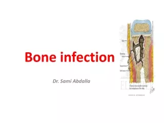
Infection of the bone and joint. Osteomyelitis. Acute osteomyelitis usually occurs in children Usually a haematogenous infection from distant focus Organisms responsible include: Staph. aureus Strep. pyogenes H. influenzae Gram-negative organisms

E N D
Osteomyelitis • Acute osteomyelitis usually occurs in children • Usually a haematogenous infection from distant focus • Organisms responsible include: • Staph. aureus • Strep. pyogenes • H. influenzae • Gram-negative organisms • Salmonella infections are often seen in those with sickle-cell anaemia • Infection usually occurs in metaphysis of long bones
Pathology • Acute inflammation results in raised intraosseous pressure and intravascular thrombosis • Suppuration produces a subperiosteal abscess that may discharge into soft tissues • Spread of infection into epiphysis can result in joint infection • Within days bone death can occur • Fragments of dead bone become separated in medullary canal (sequestrum) • New bone forms below stripped periosteum (involucrum) • If infection rapidly controlled resolution can occur • If infection poorly controlled chronic osteomyelitis can develop
Presentation • Child usually presents with pain, malaise and fever • Often unable to weight bear • Early signs of inflammation are often few • Bone is often exquisitely tender with reduced joint movement • Late infection presents with soft-tissue swellings or discharging sinus • Diagnosis can be confirmed by aspiration of pus from abscess or metaphysis • 50% of patients have positive blood cultures
Radiology • X-rays can be normal during first 3 to 5 days • In the second week radiological signs include: • Periosteal new bone formation • Patchy rarefaction of metaphysis • Metaphyseal bone destruction • In cases of diagnostic doubt bone scanning can be helpful
Management • General supportive measures should include intravenous fluids and analgesia • Painful limb often requires a splint of skin traction to relieve symptoms • Aggressive antibiotic therapy should be instituted • Flucloxacillin is often the antibiotic of choice • If fails to respond to conservative treatment surgery may be required • A subperiosteal abscess should be drained • Drilling of metaphysis is occasionally required • Overall, about 50% of children require surgery
Complications • Metastatic infection can occurs at distant sites (e.g. brain, lung) • Spread into joint can result in a septic arthritis • This complication occurs in: • Young children in whom the growth plate is permeable • Bones in which the metaphysis is intracapsular • Epiphysis of bones involved in metastatic infection • Involvement of physis can result in altered bone growth • Failure to eradicate infection can result in chronic osteomyelitis
Septic arthritis • Acute inflammatory condition of a joint • Usually results from bacterial infection • Untreated it will lead to destruction of the articular cartilage • 50% cases occur in children less than 3 years of age • In infants less than one year old the hip is the commonest joint involved • In older children the knee is the commonest joint affected • 10% of patients have multiple joints involved
Microbiology • Organism can enter joint via a number of routes • Penetrating wound • From epiphysis or metaphysis • Haematogenous spread • Provoke an acute inflammatory response • Large number of neutrophils accumulate in joint • Release proteolytic enzymes that break down the articular cartilage • Results in joint effusion and reduced synovial blood supply
Complications • Avascular necrosis of epiphysis • Joint subluxation / dislocation • Growth disturbance • Secondary osteoarthritis • Persistent or recurrent infection
Clinical features • Exact presentation depends on age • Children are usually systemically unwell • Present with pain in the affected joint • All movements of the joint are painful • Reluctant to stand on weight-bearing joints • Affected joint is usually swollen, red and warm • Hip involvement results in flexion and external rotation • In adults septic arthritis is usually associated with immunosuppression
Ix • Key investigation is culture of a joint aspirate • Should be performed prior to the administration of antibiotics • Other appropriate investigations should include • Inflammatory markers • Plain x-rays • Bone scan
DD • Irritable hip • Perthe's disease • Osteomyelitis • Gout • Pseudogout
Mx • Antibiotics should be started after joint aspiration • Empirical therapy should be commenced based on likely organisms • Adjusted depending antibiotic sensitivity • Antibiotics should be continued for 6 weeks
Sx • Involves joint drainage and lavage • May be performed arthroscopically • Early joint mobilisation should be encouraged
Pott’s disease • Pott's disease is tuberculous spondylitis • Well recognised in Egyptian mummies • Described by Percival Pott in 1779 • Now rare in western countries • Still prevalent in the developing world
Pathology • Usually occurs secondary to infection elsewhere • Due to a combination of osteomyelitis and arthritis • Often occurs at more than one vertebral level • Usually affects anterior part of vertebral body • More common in thoracic spine • Bone destruction lead to vertebral collapse and kyphosis • Spinal cord can be narrowed resulting in cord compression and neurological deficit
Clinical presentation • Back pain is the commonest symptom • Pain may be present for several months • Pain can be both spinal and radicular • 50% patients have neurological signs at presentation • Most patients have some degree of kyphosis • Cold abscess may point in the groin
Ix • Serum ESR is usually massively raised • Tuberculin skin test is usually positive • Plain x-rays may show • Lytic destruction of anterior vertebral body • Anterior vertebral collapse • Reactive sclerosis • Enlarged psoas shadow • CT or MRI provides information on disc space and neurological involvement • As allows CT guided biopsy to obtain microbiological and pathological specimens
Tx • Treatment involves both tuberculous chemotherapy and possible surgery • Nine months of combination chemotherapy should be used • This involves 3 or 4 drugs • Isoniazid and rifampicin should be given for full nine months • Pyrazinamide, ethambutol or streptomycin should be give for first 2 months • Surgery is indicated if: • Neurological deficit • Spinal deformity with instability • No response to medical treatment • Non-diagnostic percutaneous biopsy • Surgical approach depends on extent of disease and level of spinal involvement • Usually involves radical debridement and posterior stabilisation