Download
1 / 85
850 likes | 1.08k Views
Hematology . Jan Bazner-Chandler CPNP, CNS, MSN, RN. Blood. Blood is the fluid of life Blood is composed of: Plasma RBC WBC Platelets. Plasma. Plasma consists of: 90% water. 10 % solutes: albumin, electrolytes and proteins.

E N D
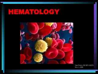
Hematology Jan Bazner-Chandler CPNP, CNS, MSN, RN
Blood • Blood is the fluid of life • Blood is composed of: • Plasma • RBC • WBC • Platelets
Plasma • Plasma consists of: • 90% water. • 10 % solutes: albumin, electrolytes and proteins. • Proteins consist of clotting factors, globulins, circulating antibodies and fibrinogen.
Red Blood Cells • RBC’s travel through the body delivering oxygen and removing waste. • RBC’s are red because they contain a protein chemical called hemoglobin which is bright red in color. • Hemoglobin contains iron, making it an excellent vehicle for transporting oxygen and carbon dioxide.
RBC’s • Average life cycle is 120 days. • The bones are continually producing new cells.
White Blood Cells • The battling blood cells. • The white blood cells are continually on the look out for signs of disease. • When a germ appears the WBC will: • Produce protective antibodies. • Surround it and devour the bacteria.
WBC’s • WBC life span is from a few days to a few weeks. • WBC’s will increase when fighting infection. • Neutropenia – abnormal percentage of neutrophils compared to total white blood cells; decreases a child’s ability to fight pathologic bacteria
Platelets • Platelets are irregularly-shaped, colorless bodies that are present in blood. • Their sticky surface lets them form clots to stop bleeding.
Blood Values • CBC with differential and platelet count. • Hgb: • Normal levels are 11 to 16 g / dl • Panic levels are: • Less than 5 g / dl • More than 20 g / dl
Hematocrit • Normal hematocrit levels are 35 to 44%. • Panic levels: • Hmct less than 15 % • Hmct greater than 60%
Blood Tests • Hemoglobin and hematocrit used to screen for anemia. • The CBC with differential would be used to help diagnose a specific disorder. • A bone marrow aspiration would be the most conclusive in determining cause of anemia – aplastic / leukemia.
Coagulation Profile • Partial thromboplastin time • Prothrombin time • Platelet count • Fibrinogen • Platelet function analysis – clotting analysis
Bone Marrow Bone marrow is the spongy substance found in the center of the bones. • It manufactures bone marrow stem cells, which in turn produce blood cells. • Red blood cells – carry oxygen to tissue • Platelets – help blood to clot • White blood cells – fight infection
Bone Marrow Transplant • Donor is placed under anesthesia. • Marrow is aspirated out of the iliac crest. • Marrow is filtered and treated to remove bits of bone and other unwanted cells and debris, transferred to a blood bag, and is infused into the patient’s blood just like at transfusion.
Treatment Modalities • Transfusion: • Packed red blood cells – anemia • Platelets – platelet dysfunction • Fresh frozen plasma – coagulation factors
Blood Transfusions • 3 types of transfusion reactions • Hemolytic • Allergic • Febrile
Hemolytic Reaction • Refers to an immune response against transfused blood cells. • Antigens, on the surface of red blood cells, are recognized as “foreign proteins” and can stimulate B lymphocytes to produce antibodies to the red blood cell antigens.
Hemolytic reaction • Flank pain • Fever • Chills • Bloody urine • Rash • Low blood pressure • Dizziness / fainting
Nursing Management • Stop the blood transfusion. • Start normal saline infusion. • Take vital signs with blood pressure • Call the MD • Obtain blood sample and urine specimen. • Return blood to blood bank. • Document
Febrile Reaction • Often occurs after multiple blood transfusions. • Symptoms:fever, chills, and diaphoresis. • Interventions: • Slow transfusion and administer antipyretic. • Administer antipyretic prior to administration.
Allergic Reaction • Symptoms: rash, urticaria, respiratory distress, or anaphylaxis. • Interventions: • administer antihistamine before transfusion • Physician may order washed rbc’s
Alteration in Hematologic Status • Disorders of hemostasis or clotting factors • Structural or quantitative abnormalities in the hemoglobin. • Anemias • Aplastic Anemia
Genetic Implications • The following have a genetic link: implications for genetic screening and fetal diagnosis • Sickle cell anemia • Thalassemia • Hemophilia
Bleeding Disorders • Three types Hemophilia: males only • Type A most common – factor VIII deficiency • Type B - lack of factor IX (Christmas Disease) • Type C – lack of factor XI Von Willebrand Disease – 1% of population – men or women – prolonged bleeding time
Hemophilia Type A • Hemophilia type A is the deficiency of clotting factor VIII. • A serious blood disorder • Affects 1 in 10,000 males in the US • Autoimmune disorder with lowered level of clotting factor • All races and socio economic groups affected equally
Hemophilia • Hemophilia is a sex-linked hereditary bleeding disorder • Transmitted on the X chromosome • Female is the carrier • Women do not suffer from the disease itself
Historical Perspective • First recorded case in Talmud Jewish text by an Arab physician – documentation of two brothers with bleeding after circumcision. • Queen Victoria is carrier and spread the disease through the male English royalty.
Goals of Care • Goals of care: • Provide factor VIII (IX) to aid blood in clotting. • To decrease transmission of infectious agents in blood products; hepatitis & AIDS. • Future: gene therapy to increase production of clotting factor.
Assessment • Circumcision may produce prolonged bleeding. • As child matures and becomes more active the incidence of bleeding due to trauma increases • May be mild, moderate or severe • Bleeding into joint spaces, hemarthrosis • Most dangerous bleed would be intracranial
Diagnosis • Presenting symptoms • Prolonged activated aPTT and decreased levels of factor VIII or IX. • Genetic testing to identify carriers
Pharmacologic Interventions • Products used to treat hemophilia are: • Fresh frozen plasma and cryoprecipitate which are from single blood donors and require special freezing • Second generation of factor VIII are made with animal or human proteins • Fibrin glue – mixture of fibrogen and thrombin can be applied topically on a wound to stop minor bleeding
Multidisciplinary Interventions • Replace the factor as ordered by physician. • Manage pain utilizing analgesics as ordered – no salicylate products. • Maintaining joint integrity during acute phase: immobilization, elevation, ice. • Physical therapy to prevent flexion contraction and to strengthen muscles and joints. • Provide opportunities for normal growth and development.
Family Education • Medic-Alert bracelet • Injury prevention appropriate for age • Signs and symptoms of internal bleeding or hemarthrosis • Dental checkups • Medication administration
Long Term Complications • 20% develop neutralizing antibodies that make replacement products less effective. • Gene therapy providing continuous production of the deficient clotting factor could be the next major advance in hemophilia treatment.
Disseminated Intravascular Coagulation or DIC • DIC is an acquired coagulopathy that is characterized by both thrombosis and hemorrhage. • DIC is not a primary disorder but occurs as a result of a variety of alterations in health.
Assessment • The most obvious clinical feature of DIC is bleeding. • Renal involvement = hematuria • Pulmonary involvement = hemoptysis, tachypnea, dyspnea and chest pain. • Cutaneous involvement = petechiae, ecchymosis, jaundice, acrocyanosis and gangrene.
Multidisciplinary Interventions • Treatment of the precipitating disorder. • Supportive care with administration of platelet concentration and fresh frozen plasma and coagulation factors. • Administration of heparin (controversial in children). • Heparin potentates anti-thrombin III which inhibits thrombin and further development of thrombosis.
Assessment • Rigorous ongoing assessment of all body systems • Monitor bleeding • No rectal temps • Avoid trauma to delicate tissue areas • All injections sites and IV sites need to be treated like an arterial stick.
Prognosis • Depends on the underlying disorder and the severity of the DIC.
ITP • Idiopathic thrombocytopenic purpura • Idiopathic = cause is unknown • Thrombocytopenic = blood does not have enough platelets • Purpura = excessive bleeding / bruising
Immune Thrombocytopenic Purpura • Antibodies destroy platelets • Antibodies see platelets as bacteria and work to eliminate them • ITP is preceded by a viral illness • URI • Varicella • Smallpox / measles vaccine • Mononucleosis • Flu
Two Types • Acute: occurs in children between 1 and 6 years • Chronic (continuous or recurrent): older than 10 years and female
Symptoms • Random purpura • Epistaxis, hematuria, hematemesis, and menorrhagia • Petechiae and hemorrhagic bullae in mouth
Diagnostic Tests • Low platelet count • Peripheral blood smear • Antiplatelet antibodies Normal platelet count: 150,000 to 400,000
Pharmacologic Interventions • For severe cases (platelet count < 20,000) • IV gamma globulin for 2-5 days to block antibody production, reduce autoimmune problem and increase platelet count • Corticosteroids to enhance vascular stability, increase platelet survival • IV anti-D to stimulate platelet production
Sickle Cell Disease • Autosomal recessive disorder • Defect in hemoglobin molecule • Cells become sickle shaped and rigid • Lose ability to adapt shape to surroundings. • Sickling may be triggered by fever and emotional or physical stress
Pathophysiology • When exposed to diminished levels of oxygen, the hemoglobin in the RBC develops a sickle or crescent shape; the cells are rigid and obstruct capillary blood flow, leading to congestion and tissue hypoxia; clinically, this hypoxia causes additional sickling and extensive infarctions.