Download
1 / 70
1.14k likes | 2.13k Views
Leukemia/Lymphoma. Components and General Properties of Blood. Seven kinds of formed elements Erythrocytes: red blood cells (RBCs) Platelets ( thrombocytes ) (clotting cells) Cell fragments from special cell in bone marrow Leukocytes: white blood cells (WBCs)-5 kinds.

E N D
Components and General Properties of Blood • Seven kinds of formed elements • Erythrocytes: red blood cells (RBCs) • Platelets (thrombocytes) (clotting cells) • Cell fragments from special cell in bone marrow • Leukocytes: white blood cells (WBCs)-5 kinds
Components and General Properties of Blood • Five leukocyte types divided into two categories • Granulocytes (with granules) • 3)Neutrophils • 4)Eosinophils • 5)Basophils • Agranulocytes (without granules) • 6)Lymphocytes • 7)Monocytes
Components and General Properties of Blood Monocyyte Small lymphocyte Neutrophil Platelets Eosinophil Small lymphocyte Erythrocyte Young (band) neutrophil Neutrophil Monocyte Large lymphocyte Neutrophil Basophil
Leukocytes • Least abundant formed element • 5,000 to 10,000 WBCs/L • Protect against infectious microorganisms and other pathogens • Conspicuous nucleus • Spend only a few hours in the bloodstream before migrating to connective tissue • Retain their organelles for protein synthesis • Granules • All WBCs have lysosomes called nonspecific (azurophilic) granules: inconspicuous so cytoplasm looks clear • Granulocytes have specific granules that contain enzymes and other chemicals employed in defense against pathogens
Types of Leukocytes • Granulocytes • Neutrophils (60% to 70%): polymorphonuclear leukocytes • Barely visible granules in cytoplasm; three- to five-lobed nucleus • Eosinophils (2% to 4%) • Large red-orange granules; • Basophils (less than 1%) • Large, abundant, violet granules
Agranulocytes • Lymphocytes (25% to 33%) • Variable amounts of bluish cytoplasm (scanty to abundant); ovoid/round, uniform dark violet nucleus • Monocytes (3% to 8%) • Largest WBC; generally ovoid, kidney-, or horseshoe-shaped nucleus
Granulocytes • Neutrophils—increased numbers in bacterial infections • Phagocytosis of bacteria • Release antimicrobial chemicals • Eosinophils—increased numbers in parasitic infections, collagen diseases, allergies, diseases of spleen and CNS • Phagocytosis of antigen–antibody complexes, allergens, and inflammatory chemicals • Release enzymes to destroy large parasites
Basophils—increased numbers in chickenpox, sinusitis, diabetes • Secrete histamine (vasodilator): speeds flow of blood to an injured area • Secrete heparin (anticoagulant): promotes the mobility of other WBCs in the area
Agranulocytes • Lymphocytes—increased numbers in diverse infections and immune responses • Destroy cells (cancer, foreign, and virally infected cells) • “Present” antigens to activate other immune cells • Coordinate actions of other immune cells • Secrete antibodies and provide immune memory
Agranulocytes • Monocytes—increased numbers in viral infections and inflammation • Leave bloodstream and transform into macrophages • Phagocytize pathogens and debris • “Present” antigens to activate other immune cells—antigen-presenting cells (APCs)
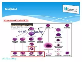
The Leukocyte Life Cycle • Leukopoiesis—production of white blood cells • Pluripotent stem cells (PPSCs) • Myeloblasts—form neutrophils, eosinophils, basophils • Monoblasts—form monocytes • Lymphoblasts give rise to all forms of lymphocytes
Leukopoiesis Mature cells Pluripotent stem cell Colony-forming units (CFUs) Precursor cells leaves Eosinophilic CFU Eosinophilic myeloblast Eosinophilic promyelocyte Eosinophilic myelocyte Eosinophil Basophilic CFU Basophilic myeloblast Basophilic promyelocyte Basophilic myelocyte Basophil Neutrophilic CFU Neutrophilic myeloblast Neutrophilic promyelocyte Neutrophilic myelocyte Neutrophil Monocytic CFU Monoblast Promonocyte Monocyte B lymphocyte B prolymphocyte T lymphocyte T prolymphocyte Lymphocytic CFU Lymphoblast NK cell NK prolymphocyte
What Is Leukemia? • Cancer of the white blood cells • Acute or Chronic • Affects ability to produce normal blood cells • Bone marrow makes abnormally large number of immature white blood cells called blasts
History • Means “white blood” in Greek • Discovered by Dr. Alfred Velpeau in France, 1827 • Named by pathologist Rudolf Virchow in Germany, 1845
Leukocyte Disorders • Leukemia—cancer of hemopoietic tissue that usually produces an extraordinary high number of circulating leukocytes and their precursors • Myeloid leukemia: uncontrolled granulocyte production • Lymphoid leukemia: uncontrolled lymphocyte or monocyte production
Acute vs Chronic Leukemia • Acute leukemia: appears suddenly, progresses rapidly, death within months –blasts found in peripheral blood • Chronic leukemia: undetected for months, survival time average of 3 years • Effects: normal cell percentages disrupted; impaired clotting; opportunistic infections
Main Types • Acute Lymphocytic Leukemia (ALL) • Acute Myelogenous Leukemia (AML) • Chronic Lymphocytic Leukemia (CLL) • Chronic Myelogenous Leukemia (CML)
Demographics of Leukemia Patients (2001 Data) CLL=Chronic Lymphocytic ALL=Acute Lymphocytic CML=Chronic Mylogenous AML=Acute Mylogenous Total Reported Cases = 31,500
Platelet Platelet White Cell Red Cell Red Cell Blasts White Cell Normal human blood Blood with leukemia Pictures Of Blood
Stage 1- Normal Stage 2- Symptoms Stage 3- Diagnosis Stage 5a- Anemia Stage 4- Worsening Stage 5b- Infection Development of Leukemia in the Bloodstream Legend White Cell Red Cell Platelet Blast Germ
Causes • High level radiation/toxin exposure • Viruses • Genes • Chemicals • Mostly unknown
Signs and Symptoms of AML • Insidious nonspecific onset • Pallor due to anemia • Febrile (fever) due to ineffective WBC • Petechiae (skin bruising) due to thrombocytopenia • Bone pain Petechiae
Typical Labs of AML • Leukocytosis • Blastemia • Leukemic hiatus • Auer rods – only found in myelocytic blasts • Thrombocytopenia • Anemia • >20% blasts in Bone Marrow
Auer Rods Auer Rod
CD Markers • The cluster of differentiation (cluster of designation) (often abbreviated as CD) is a protocol used for the identification and investigation of cell surface molecules providing targets for immunophenotyping of cells. • The CD markers can be used to identify the type of cell.
Other Findings • CD 13 and CD 33 in flowcytometry • Cytochemistries-stains that can be used to differentiate leukemias • Myeloperoxidase • Sudan black B • Choloroacetate esterase (specific) • Nonspecific esterase
Flow Cytometry Large clustering of CD 33s shows presence of blasts
FAB (1976) Classification • M0 -- Undifferentiated AML • M1 -- AML without maturation • M2 -- AML with maturation • M3 -- Acute Promyelocytic Leukemia • M4 -- Acute Myelomonocytic Leukemia • M5 -- Acute Monocytic Leukemia • M6 -- Erythroleukemia (DiGuglielmo’s) • M7 -- Megakaryoblastic Leukemia
Myeloperoxidase (MPO) p-Phenylene diamine + Catecol + H2O2 MPO > Brown black deposits Brown deposits considered to be a positive test-differentiates AML from other leukemias
M3 M4 M5
Chloracetate (Specific) Esterase Myeloid Cell Line Naphthol-ASD-chloracetate CAE > Free naphthol compounds + Stable diazonium salt (eg, Fast Corinth) > Red deposit
Non-Specific Esterase Monocytic Line Naphthyl acetate ANAE > Free naphthyl compounds +Stable diazonium salt (eg, Fast blue RR) > Brown deposits
FAB criteria Morphology Cytochemistry WHO criteria Morphology Immunophenotyping Genetic features Karyotyping Molecular testing Clinical features FAB vs WHO Classifications of Hematologic Neoplasm
WHO Classification of AML • AML with recurrent cytogenic translocations • AML with multi-lineage dysplasia • AML and myelodysplasia, therapy related • AML, not otherwise categorized
AML with Recurrent Cytogenetic Translocations (WHO 1995) • t(8;21) -- some maturation of neutrophilic line; rare in older patients; AML1/ETO fusion protein; >90% FAB M2 • t(15;17) -- APL (granular and microgranular variants); retinoic acid receptor (RAR) leukemias; middle aged adults; DIC • inv(16) or t(16;16) -- monocytic and granulocytic; abnormal eosinophilic component • 11q23 -- monocytic; children; most common is t(9;11)
Lymphocytic Leukemias • Can involve T or B lymphocytes • B lymphs mature in bone marrow • Responsible for making antibodies • T lymphs go to thymus to mature • Are cytotoxic cells of immune system
FAB Classification of ALL • L1: Small homogeneous blasts; mostly in children • L2: Large heterogeneous blasts; mostly in adults • L3: “Burkitt” large basophilic B-cell blasts with vacuoles
L2 L3
Periodic Acid Schiff Periodic acid + Glycogen oxidation > Aldehyde + Schiff reagent (para-rosaniline, Na metabisulfite) > Red deposit
ALL Cytochemistries • Oil Red O: stains L3 vacuoles • Terminal deoxynucleotidyltransferase (Tdt): DNA polymerase in early lymphoblasts • Cell surface markers (CD’s) • Cytoplasmic and surface immunoglobulins: B-cell line • T-cell receptor (TCR)
WHO Classification of Lymphoproliferative Syndromes • Precursor B Lymphoblastic Leukemia/Lymphoma (ALL/LBL) -- ALL in children (80-85% of childhood ALL); LBL in young adults and rare; FAB L1 or L2 blast morphology • Precursor T ALL/LBL -- 15% of childhood ALL and 25% of adult ALL • Burkitt Leukemia/Lymphoma (FAB L3)