Download
1 / 28
280 likes | 411 Views
Nervous System Review. How is the nervous system divided? . Structure The directional flow of information Control of effectors Organ, gland, or muscle that responds to a nerve stimulus. Nervous system tissue is divided by location. Central Nervous System (CNS)

E N D
How is the nervous system divided? • Structure • The directional flow of information • Control of effectors • Organ, gland, or muscle that responds to a nerve stimulus
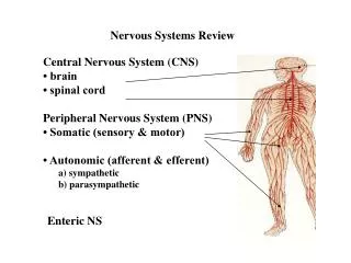
Nervous system tissue is divided by location • Central Nervous System (CNS) • Includes brain and the spinal cord • Peripheral Nervous System (PNS) • includes the nerves and their branches
Afferent and Efferent Divisions • Tissue of both the CNS and PNS have nerve cells that form incoming and outgoing pathways for information • Afferent division- includes incoming sensory pathways (nerves that carry impulses toward the CNS) • Efferent division- include outgoing motor pathways (nerves that carry impulses away from CNS to glands or muscles)
Somatic and Autonomic Nervous systems division (by the type of effectors they regulate) • Somatic Nervous system SNS Motor neurons that control voluntary actions of the skeletal muscles (Somatic effectors) • Autonomic Nervous System (ANS) Neurons that regulate involuntary actions
Somatic sensory division Cont. • SNS includes afferent pathways that provide feedback from the somatic effectors • SNS includes the integrating centers that receive the sensory information and generate the efferent response signal
Autonomic Nervous systems Cont. • Pathways carry information to the autonomic or visceral effectors.(smooth muscle, cardiac and glands) • Two efferent pathways • Sympathetic division – paths that exit middle of the spinal cord ( deal with immediate threat to internal environment) ‘flight or fight’ response • Parasympathetic division- paths that exit at the brain or lower spinal cord (deal with normal resting activities) “rest and repair” division
Cells of the Nervous System • Neurons- nerve cell including process (axons and dendrites) • Glia or Neuroglia –nonexcitable supporting cells of nervous tissue ( 5 major types to know)
Neuroglia Cells • Astrocytes – “star” largest and most numerous, only in CNS, “feed” the neurons by picking up glucose from the blood, converting it to lactic acid, and passing it to neurons • Microglia- small, stationary, CNS. In inflamed or degeneratin brain tissue cell enlarge and carry on phagocytosis • Ependymal Cells- resemble epithelial cells, form thin sheets, line fluid-filled cavities in the brain and spinal cord. Some have cillia to keep fluid moving • Oligodendrocytes – “cell with few branches” some cluster around nerve cell bodiees some are in rows between nerve fibers. Help hold nerve fibers together, produce the fatty myelin sheath in CNS • Schwann cells- Only in PNS (serve the same role as Oligodendrocytes in CNS) support nerve fibers and form mylein sheath
Schwann Cells cont. • Myelin – white fatty substance the sheath is made of • Nodes of Ranvier – gaps in the sheath between schwann cells. Important for proper conduction of impulses in PNS • Neurilemma – “sheath of Schwann” Cells nucleus and cytoplasm squeezed to the perimeter. Essential to the regeneration of injured nerve fibers (Lorenzo)
Neurons • Consist of cell body (soma, perikaryon) • At least two processes- one axon and two or more dendrites. ( extensions of cell body called nerve fibers) • Axons – conduct impulses away from the cell body. End in branches called telodendria terminating in synaptic Knob. Can be myelinated (affects the speed of impulse) • Dendrites- receive stimuli and conduct electrical signals toward the cell body (never myelinated) • Cytoplasm contains neurofibrils and neurofilaments
Neuron Classification • According to the number of their extensions from cell body • Three Types • 1) Multipolar- One axon several dendrites. Most in CNS are multipolar • 2) Bipolar- One axon and one highly branched dendrite. In the eye, inner ear, and olfactory pathway • 3) Unipolar- Are always sensory neurons conducting information to the CNS. Single process from cell body branches to form a central process to CNS and peripheral process away from the CNS (forms the axon)
Neuron Classification • According to their function • Afferent neuron- (sensory) transmit nerveimpulses to the spinal cord or brain • Efferent neuron- (motor) transmit nerve impulses away from the brain or spinal cord to muscles • Interneurons- lie entirely within the CNS, transmit impulses from afferent neurons to motor neurons
Nerves and Tracts • Nerves- bundles of peripheral nerve fibers surrounded by connective tissue • Endoneurium- surrounds each nerve fiber • Perineurium- surround bundles of endoneurium covered fibers called fascicles • Epineurium- surround bundles of Perineurium and blood vessels • Tracts- Within CNS nerve fibers are called Tracts rather than nerves
Nerve impulses • Difference in concentration of ions across the plasma membrane creating a wave of electrical fluctuations • Membrane potential is polarized both + and- • Resting membrane potential (RMP)- nonconducting neuron’s plasma membrane • Can be maintained as long as its sodium-potassium pump operate and its permeability remain stable
Impulse cont. • Local Potentials (graded potentials)- potentials above or below RMP in response to a stimuli. Dependant on magnitude of the stimulus. Excitation- occurs when a stimulus triggers the opining of stimulus-gated Na+ channels. Action Potential- Neuron that is conducting an impulse
Two Protective coverings of brain and spinal cord • Cranial bones-outer coverings • Meninges-Inner coverings (3 layers) • Dura Matter- strong, white fibrous tissue; outer layer of meninges and inner periosteum of the cranial bone • Arachnoid membrane- layer between dura mater and pia mater • Pia mater- innermost, transparent layer contains blood vessels
The Brain (six divisions) • Medulla oblongata- lowest part of the brainstem, enlarged extension of spinal cord, contains vital centers • Pons- between medulla oblongata and midbrain, composed of white matter tracts • Midbrain- conducts impulses between the midbrain and cerebrum
Cerebellum-second largest part of brain, composed of white matter called (arbor vitae) functions with the cerebrum to produce skilled skeletal muscle movements. Maintains posture and equilibrium (balance) • Diencephalon- includes • thalamus (auditory & visual input), • hypothalamus,( regulates appetite, body temperature, emotions). • pineal body(regulates body’s biological clock, produces hormones (melatonin)
Cerebrum largest division of brain • Right and Left hemisperes divided into five parts • Cerebral cortex- outer surface gray matter • Gyri- convolutions of the brain • Sulci- shallow grooves • Fissures- deeper grooves, divide cerebral hemispheres into lobes
Functions of cerebral cortex • Functional areas- one function ex. Receptors to temp. or stimulate skeletal muscle, auditory, or visual areas • Sensory functions- intetgrates pieces into whole perceptions • Motor functions- controls individual muscles or groups • Integrative functions-Consciousness, Language, Emotions, Memory • Specialized cerebral hemispheres • Electroencephalogram
Integrated functions broke down • Consciousness – state of awareness of oneself • Language- ability to speak and write • Speech center in the frontal, parietal, and temporal lobes • Left cerebral hemisphere contains speech center in 90% of population • Emotions • Limbic system known as the “emotional brain” Example anger, fear sadness, joy etc. • Memory- storing and retrieving both short and long term memory
Brain Stem • Made up of three parts • Medulla Oblongata • Midbrain • Pons • Functions • Medulla Oblongata- contains vital reflex centers cardiac, vasomotor, and respiratory. Non-vital reflex centers vomiting, coughing, sneezing, etc. • Midbrain-
Spinal Nerves • Overview • Thirty-one pairs of spinal nerves are connected to the spinal cord (Figure 14-1) • No special names; are numbered by level of vertebral column at which they emerge from the spinal cavity • Eight cervical nerve pairs (C1 through C8) • Twelve thoracic nerve pairs (T1 through T12) • Five lumbar nerve pairs (L1 through L5) • Five sacral nerve pairs (S1 through S5) • One coccygeal nerve pair • Lumbar, sacral, and coccygeal nerve roots descend from point of origin to lower end of spinal cord (level of first lumbar vertebra) before reaching the intervertebral foramina of the respective vertebrae, through which the nerves emerge • Cauda equina—describes the appearance of the lower end of the spinal cord and its spinal nerves as a horse’s tail
Spinal Nerves • Structure of spinal nerves • Each spinal nerve attaches to spinal cord by a ventral (anterior) root and a dorsal (posterior) root • Dorsal root ganglion—swelling in the dorsal root of each spinal nerve • All spinal nerves are mixed nerves
PNS • Somatic Motor Nervous System • Includes all the voluntary motor pathways outside the CNS • Involve the peripheral pathways to the skeletal muscles, which are the somatic effector • Somatic reflexes- predictable response to a stimuli
Important Somatic Reflexes • Knee Jerk Reflex- etension of the lower lef in response to tapping of the patellar tendon • Ankle Jerk Reflex- Extension of the foot in response to tapping of the Achilles tendon. • Babinski Reflex- Extension of the great toe, with or without fanning of the other toes. Normal for infants up to 1 ½ years of age then becomes plantar flexion • Corneal Reflex- blinking in response to touching the cornea. • Abdominal Reflex- drawing in of the abdominal wall in response to stroking the side of the abdomen