Download
1 / 23
390 likes | 2.81k Views
CALCIUM IMAGING. Beata M. Wolska, Ph.D. Department of Medicine, Section of Cardiology & Department of Physiology & Biophysics University of Illinois at Chicago October 16, 2001. INTRODUCTION. Ca 2+ is a universal second messenger

E N D
CALCIUM IMAGING Beata M. Wolska, Ph.D. Department of Medicine, Section of Cardiology & Department of Physiology & Biophysics University of Illinois at Chicago October 16, 2001
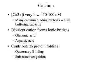
INTRODUCTION • Ca2+ is a universal second messenger • Ca2+ is involved in specific & selective regulation of cellular processes (muscle contraction, fertilization, synaptic transmission, cell division, blood clotting etc.) • Changes in intracellular Ca2+ concentration are often within ms • Ca2+ can not be visualized directly in living cells • To image changes in Ca2+ concentration specific molecules are used that have optical properties, which change upon interacting with Ca2+
FLUORESCENCE TECHNIQUES(THE FLUORESCENCE PROCESS) • Fluorescence is the result of a three-stage process that occurs in molecules called fluorophores or fluorescent dyes • Stages of fluorescence • Stage 1 – Excitation • Stage 2 – Excited-State Lifetime • Stage 3 – Fluorescence Emission • The process responsible for the fluorescence of fluorescent dye can be schematically illustrated using the electronic-state diagramcalled the Jablonski diagram
JABLONSKI DIAGRAM Singlet excited state Excited electronic singlet state Excited-State Lifetime (10-9-10-8 s) Excitation Fluorescence Emission Stokes shift huEX - huEM Ground state FLUORESCENCE QUANTUM YIELD # fluorescence photons emitted (Stage 3) # fluorescence photons absorbed (Stage 1)
FLUORESCENCE DETECTION • FLUORESCENCE INSTRUMENTATION • FLUORESCENCE SIGNALS • BACKGROUND FLUORESCENCE • MULTICOLOR LABELING EXPERIMENTS • RADIOMETRIC MEASUREMENTS
FLUORESCENCE INSTRUMENTATION Fluorescence detection system 1) Fluorophore 2) Wavelength filters 3) Detector 4) Excitation source Types of fluorescence instruments 1) Spectrofluorometer 2) Fluorescence microscope 3) Flow cytometer
SELECTION CRITERIA FOR Ca2+ INDICATORS • Ca2+concentration range of interest (dissociation constant Kd; detectable response 0.1Kd to 10Kd) • The method of delivery of the indicator to the cell • Measurement mode (quantitative vs. qualitative ion concentration data, type of instrument, source of noise etc.) • The intensity of the light emitted from the indicator • Simultaneous recordings of other physiological parameters
SCHEMATIC DIAGRAM OF LOADING THE CELLS USING ACETOXYMETHYL (AM) ESTER DERIVATIVE FURA-2/AM PROBLEMS: Compartmentalization Incomplete AM ester hydrolysis Leakage
FLUORESCENT Ca2+ INDICATORS EXCITED WITH UV LIGHT • Fura-2, Indo-1 and derivatives • Quin-2 and derivatives • Indicators with intermediate calcium-binding affinity (Fura-4F, Fura-5F & Fura-6F; Benzothiaza-1 & Benzothiaza-2) • Low-affinity calcium indicators (Fura-FF, BTC, Mag-Fura-2, Mag-Fura-5, Mag-Indo-1)
FLUORESCENCE EXCITATION SPECTRA FURA-2 Kd~135 nM (Mg2+-free) Kd~224 nM (Mg2+ 1mM) BIS-FURA-2 Kd~370 nM (Mg2+-free) Kd~525 nM (Mg2+ 1mM)
FLUORESCENT Ca2+ INDICATORS EXCITED WITH VISIBLE LIGHT - ADVANTAGES • Efficient excitation with most laser-based instrumentation, including confocal laser scanning microscope • Reduced interference from sample autofluorescence • Less cellular photodamage and light scatter • Higher absorbance of the dye • Compatibility with UV probes and “caged” probes
FLUORESCENT Ca2+ INDICATORS EXCITED WITH VISIBLE LIGHT • Fluo-3, Rhod-2 and related derivatives • Low-affinity calcium indicators: Fluo-5N, Rhod-5N, X-Rhod-5N and related derivatives • Calcium Green, Calcium Orange and Calcium Crimson indicators • Oregon Green 488 BAPTA indicators • Fura Red indicator • Calcein
FLUORESCENCE EMISSION SPECTRA OF THE MIXTURE OF FLUO-3 & FURA RED INDICATORS
RATIOMETRIC CALIBRATION • Used only when dyes show an excitation or emission spectral shift upon ion binding • Fluorescence intensities are measured at two different wavelengths (with opposite ion-selective responses) • Radiometric measurements reduce variations of several factors including indicator concentration, excitation pathlength, excitation intensity, detector efficiency
EXAMPLE OF FURA-2 RATIO CALIBRATION USING A Ca2+ IONOPHORE [Ca]=Kd x (R-Rmin)/(R-Rmax) x Sf2/Sb2 Rmin=A1/A2 Rmax=B1/B2 Sf2/Sb2=A2/B2
FLUORESCENT Ca2+ INDICATOR CONJUGATES • Dextran conjugates • Fura and Indo Dextrans • Calcium Green and Oregon Green 488 BAPTA Dextrans • Lipophilic derivatives for detecting near-membrane calcium • Fura-C18 and Calcium Green-C18
A BIOLUMINESCENT Ca2+ INDICATOR • Production of light by biological organisms (photoproteins) is called bioluminescence • Bioluminescence does not require illumination • Intensity of of light produced is often low • Assays based on bioluminescence are sensitive and free of background • Aequorin is a photoprotein isolated from luminescent jellyfish • Aequorin is not exported or secreted, it is not compartmentalized and is typically microinjected into cells
FURA-2 FLUORESCENCE RATIO OF MOUSE MYOCYTES O MIN 40 MIN 2.5 2.0 FURA-2 RATIO (340/380) 1.5 2 SEC 1.0