Download
1 / 58
710 likes | 1.68k Views
Esophageal Emergencies. Tintinalli chapter 75. Anatomy/Physiology. Muscular tube 20-25 cm long Majority in mediastinum, post/lat to trachea Outer longitudinal & inner circular muscles Upper 1/3 striated muscle, lower half all smooth muscle

E N D
Esophageal Emergencies Tintinalli chapter 75
Anatomy/Physiology • Muscular tube 20-25 cm long • Majority in mediastinum, post/lat to trachea • Outer longitudinal & inner circular muscles • Upper 1/3 striated muscle, lower half all smooth muscle • Two sphincters; UES cricopharyngeus muscle, LES lower 1-2 cm of esophagus
Anatomy/Physiology • Three anatomic constrictions: • Cricopharyneus • Aortic arch/left mainstem bronchus • Gastroesophageal junction
Anatomy/Physiology • Innervation mirrors cardiac, a convergence of somatic and visceral stimuli; cardiac and esophageal chest pain similar. • Blood supply; inferior thyroid artery, branches from thoracic aorta, branches from left gastric and inferior phrenic arteries.
Anatomy/Physiology • Venous; submucosal plexus drains into plexus outside of esophagus • Outer plexus to : • Inferior thyroid • Azygos • Coronary • Gastric venous system
Dysphagia • Defined; difficulty swallowing • Majority will have organic process • Two types: • Transfer dysphagia; early in swallowing process • Transport dysphagia; impaired movement down esophagus through LES, perceived later in process, feeling of food “getting struck”
Dysphagia • Transfer: • 80 % neuromuscular; CVA,scleroderma, myasthenia gravis, parkinsons, lead poisoning, thyroid disease • Risk of aspiration • Discoordinated food bolus transfer to esophagus • Symptoms; gagging, coughing, nasal regurg.
Dysphagia • Transport: • 85 % obstructive disease; foreign body, carcinoma, webs, strictures, thyroid enlargement, vessel abnormalities, diverticuli • Less aspiration risk • Improper transfer from esophagus to stomach • symptoms; food sticking, retrosternal fullness, odonophagia
Dysphagia • History • Acute vs. chronic • Solids vs. liquids • Intermitent or progressive • Feeling of “something stuck” • Physical exam; focus on head and neck and neuro, helpful to watch pt swallow sip of water. Physical exam often normal.
Dysphagia • ED work-up: AP & lat neck x-rays. CXR. • Diagnosis often made outside ED. Barium swallow usually first test. Ultimately best worked up with video-esophagography.
Structural/Obstructive causes • Neoplasm: common cause of both types. • 95 % squamous cell • Male : female , 3:1 • Fast progression from solids to liquid dysphagia • Pts >40 yo with dysphagia assume neoplasm. Need expedient work up to rule out malignancy
Structural/Obstructive causes • Esophageal stricture: results from scaring from GERD • Generally distal esophagus, may interfere with LES • Symptoms develop over years, usually only solids • Must rule out malignancy
Structural/Obstructive causes • Schatzki ring: most common cause of intermittent dysphagia with solids • Fibrous stricture near GE junction in 15 % of population • Pts frequently present with food impacted after poorly chewed meat • Treatment is dilatation
Structural/Obstructive causes • Esophageal webs: thin structures of mucosa and submucosa • Often mid or proximal esophagus • Congenital or acquired • Plummer-Vinson syndrome, with iron deficiency anemia • Tx is dilatation
Structural/Obstructive causes • Diverticula: can be found throughout esaphagus • Zenker; progressive outpouching of pharyngeal mucosa above UES. d/t increased pressure when swallowing. • Usually seen after age 50 • Halitosis • Feeling of a neck mass
Motor lesion causes • Neuromuscular disorders; misdirection of food bolus, • liquid > solids. • Symptoms intermittent. • CVA #1 cause • Polymyositis/Dermatomyositis #2 causes
Motor lesion causes • Achalasia; dysmotility disorder, • unknown cause. • Impaired LES relaxation, • absence of esophageal peristalsis. • Patients 20-40 yo. • Symptoms; regurgitation, weight loss, odonophagia
Motor lesion causes • Diffuse esophageal spasm; • dysphagia intermittent and does not progress. • Tx =control any reflux present
Chest Pain of Esophageal Origin • Differentiating esophageal from ischemic pain very difficult. Often not done in ED. • Pain at night, spontaneous onset, regurgitation, odynophagia, dysphagia, meal induced= can be seen in both • High admission rate of chest pain found not to be cardiac is appropiate. • 20-60% of chest pain is esophageal and normal coronary arteries.
GERD • 25% of adults • Weak or transient relaxing of LES is primary cause • Other causes= high fat diet, nicotine, ETOH, caffeine, pregnancy, meds(nitrates, Ca channel blockers, estrogen, progesterone) • Heartburn is classic symptom
GERD • Other symptoms= odynophagia,dysphagia, acid regurgitation, hyperslivation. Asthma exac, dental erosions, frequent URI’s, vocal cord ulcers, laryngitis, hoarseness, chronic cough • Postural changes in pain= increasing intraabdominal pressure can increase pain • Relief with antacids
GERD • Complications= strictures, esophageal inflammation, Barrett esophagus (columnar epith replaces strat squamous) premalignant. • Pain; squeezing, pressusre-like, onset with exertion, diaphoresis, pallor, nausea, vomiting, radiation to arms and neck, shoulder and back. All similar to cardiac pain
GERD • TX; • Avoid exacerbating agents • Elevate head of bed 30 degrees • Don’t eat 3 hours before going to bed • H2 blockers or PPI’s
Esophagitis • Inflammatory: can progress to ulceration, scarring, stricture • Reflux induced- aggressive tx. with acid suppression • Med induced-NSAIDs, KCL, doxy, clinamycin, tetracycline • Infectious: immunosuppression; AIDS • Candida #1, HSV, CMV, aphthous ulceration • Fungal, vericella, EBV.
Esophageal Motility Disorders • Chest pain, dull/achy, at rest, 5th decades, intermittent dysphagia • Esophageal dysmotility: excessive, uncoordinated contraction • Achalasia & diffuse es. spasm as above • Ineffective esophageal motility • Hypertensive LES • Nutcracker esophagus; high amplitude, long duration contractions LES, >180 mmHg • Tx with NTG, Ca channel blockers
Esophageal Perforation • Iatrogenic 75% • endoscopy #1 cause • Boerhaave syndrome 10-15% • ETOH • emesis
Esophageal Perforation • Trauma 10% • Blunt rare • Penetrating wounds more common, often masked by more critical wounds in the area • FB ingestion; perforation usually at anatomic narrowings. d/t pressure necrosis(coin), penetrating from sharp object(pin), chemical irritant(battery)
Esophageal Perforation • Esophageal contents to ; • Mediastinum-fulminant necrotizing mediastinitis and polymicrobial infection to shock • Pleural/peritoneal space- rapidly progressive infection/shock • Most spontaneous perfs through left post-lat wall in distal esophagus. Proximal perfs with instrumentation
Esophageal Perforation • Pain- acute, severe, unrelenting, diffuse, chest neck and abdomen. • May radiate to back and shoulders • Exacerbated by swallowing • Dysphagia, dyspnea, hematemesis, cyanosis may all be present • Confused w/(MI, PE, ulcer, aortic catastrophe, acute abd.)= delays in dx.
Esophageal Perforation • Physical; abd rigidity, hypotension, fever, tachycardia, tachypnea all common. • Cervical sub-q emphysema if cervical perf • Mediastinal emphysema takes longer • “Hammon crunch” air in mediastinum being moved by beating heart • Pleural effusion in 50% w/ intrathoracic perfs.
Esophageal Perforation • CXR=suggestive • CT=confirms • Endoscopy= confirms • Pleural fluid aspirate= high amylase
Esophageal Perforation • High mortality rate regardless of cause • Location, etiology, time until dx all affect outcome • Rapid aggressive mgt is key • Tx shock • Surgical consult
Esophageal Bleeding • General approach: • UGIB= airway mgt., NG tube, gastric lavage, blood if needed, GI consult • 60% vericeal bleeds resolve w/ supportive care. 80% if bleed is not vericeal. • If continue to bleed= early endoscopy • Pharmacologic= somatostatin analogs • Balloon tamponade= last resort
Esophageal Bleeding • Varices: • Seen in chronic liver ds & portal HTN • 60% of pts with chronic liver ds. • 25-30% experience hemorrhage • Varices from ETOH abuse have higher risk of bleeding • 2/3 that bleed have recurrent hemorrhage
Esophageal Bleeding • Varices: • Endoscopy first line to control bleeding • Sclerotherapy and ligation are alternatives • Despite tx, mortality remains high
Esophageal Bleeding • Mallory-Weiss syndrome: • Arterial bleeding from longitudinal mucosal lacs. of distal esoph/prox stomach • 5-15% of UGIB • 4th – 6th decades • Acute onset of UGIB • Overall low relative incidence of surgical intervention or adverse outcomes is seen
Esophageal Bleeding • Mallory-Weiss syndrome: • Initial tx = supportive, most stop spontaneously • Ongoing bleeding= electrocoagulation, sclerotherapy, laser photocoagulation, angiographic embolization, surgery.
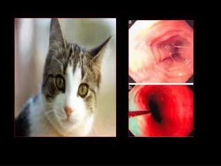
Esophageal Bleeding • Esophageal Cancer: • Heme-positive stools • Uncommon cause of significant UGIB or LGIB
Swallowed Foreign Bodies Tintinalli Chapter 76
Swallowed FB • Peds 80% of all cases • Prisoners, psych, edentulous adults • Adults=meat and bones • Peds = coins, toys, crayons, pen caps • Psych and prisoners = unlikely objects, spoons, razors
Pathophysiology • Most pass spontaneously • 10-20% require some intervention • 1% surgical • Most are at “anatomic narrowings” • Peds: cricopharyngeal(C6) most common, thoracic inlet(T1), aortic arch(T4), tracheal bifurcation(T6), hiatal narrowing(T10-11)
Pathophysiology • Once object passes pylorus, usually passes out with stool. • Irregular or sharp edges may lodge anywhere though.
Clinical Presentation • Objects in esophagus: • Anxiety, discomfort, retrosternal pain, retching, vomiting, dysphagia, choking, coughing. • In peds= refusal to eat, vomiting, gagging, choking, stridor, neck or throat pain, increased salivation, inability to swallow.
Clinical Presentation • Physical exam: • Nasopharynx, oropharynx, sub-q tissue for air. • Laryngoscopy (direct or indirect) • Objects warrenting endoscopy consult: • Sharp/elongated, multiple FB, button batteries, evidence of perf, child w/ coin at cricopharyngeous, airway compromise, FB for >24 hours
ED Management • General Care: • Expectant once FB past pylorus • If FB obstructs esoph, insert tube above FB to remove unswallowed material • Locate FB: • Standard x-ray • Endoscopy= locates and removes FB, procedure of choice • Esophagogram- consult endoscopist prior to contrast
ED Management • Type of contrast: • Perf expected= water soluble contrast, Gastrografin • Aspiration is possible use Barium, Gastrografin is pulmonary irritant • Perf and aspiration possible: use nonionic contrast
ED Management • Monitor FB progress w/ x-rays 2-4 hrs apart • Frequent abd exams for peritonitis should perf occcur
ED Management • Food impaction: • Meat= time and sedation allow meat to pass. Do not allow in esoph >12 hrs • Endoscopy #1 • Glucagon 1 mg IV, repeat 2 mg IV in 20 min, relaxes esoph smooth muscle • Nifedipine 10 mg sub lingual, reduces LES pressure • DO NOT use meat tenderizer d/t complications including perforation
ED Management • Coin injestion: (usually children) • 35% are asymptomatic • Coins lie in frontal plane in esoph, = flat side visible on AP films • Coins in trachea in sagittal plane • Foley catheter removal if <24 hrs • Secondary to endoscopy • Protect airway first = ET tube
ED Management • Button Battery: • True emergency, rapid action of alkaline on mucusa, burns in 4 hrs, perfs in 6hrs. • Lithium cells= bad outcomes • Mercury containing= get blood and urine mercury levels