Download
1 / 122
1.23k likes | 1.75k Views
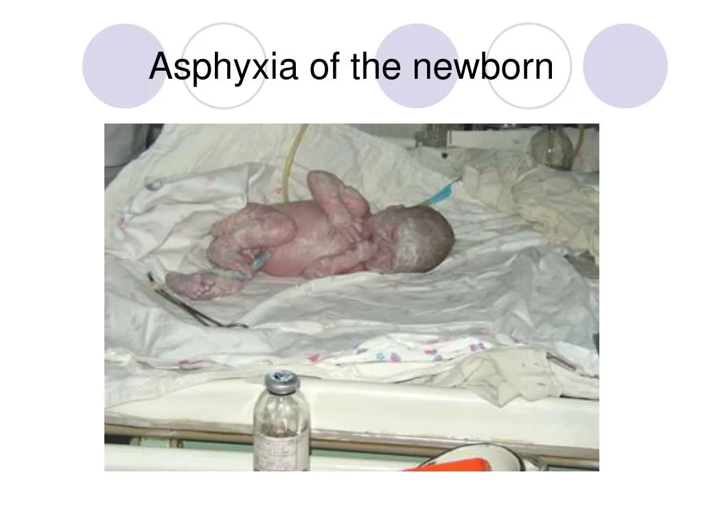
ASPHYXIA OF NEWBORN IN NICU PEDIATRICS <br>

E N D
Definition • WHO: Asphyxia is incapacity of newborn to begin or to support of spontaneous respiration after delivery due to breaching of oxygenation during labor and delivery • India: Asphyxia is absent or ineffective respiration of newborn of 1 minute old with Apgar score less than 4
Definition • Great Britain: Asphyxia is critical insufficiency of oxygen in fetus during delivery so severe that leads to development of metabolic acidosis and depression of spontaneous respiration
Definition • Canada: Asphyxia is breach of gas exchange when hypoxia and hypercapnia, and considerable metabolic acidosis occur
Definition • Australia: Asphyxia is a state with • mother has complications in perinatal period that decrease provision with oxygen and leads to acidosis • functional violation minimum 2 organs due to acts of acute hypoxia
Definition • Ukraine: Asphyxia of newborn as a nosological form is conditioned by causes when fetus out and find (connect) with severe maternal-placental and (or) umbilical flow leads to increasing of oxygen approach to fetus tissue and hypoxia development
Asphyxia: means to be pulse less, but more useful is a definition of impaired or interrupted gas exchange. These situations can take place: a. Intrauterine: the gas exchange depends on the function of placenta, and the blood-flow in the umbilical vessels. b. Intrapartum c. Postnatal: after delivery the gas exchange takes place in the pulmonary vesicles or alveoli and depends on the function of the heart, lungs and brain. Asphyxia
Causes of Asphyxia • Fetal hypoxia: • Mother: hypoventilation during anesthesia, cyanotic heart disease, respiratory failure or carbon monoxide poisoning. • Low maternal blood pressure as a result of the hypotension that may compression of the vena cava & aorta by the gravid uterus • Inadequate relaxation of the uterus to permit placental filling as a result of uterine tetany caused by excessive administration of oxytocin • Premature separation of the placenta; placenta previa • Impedance to the circulation of blood through the umbilical cord as a result of compression or knotting of the cord • Uterine vessel vasoconstriction by cocaine, smoking • Placental insufficiency from numerous causes, including gestosis, eclampcia, toxemia, postmaturity • Extremes in maternal age (< 20 years or >35 years) • Preterm or postterm gestation.
Causes of Asphyxia • Intrapartus asphyxia: • More frequently inadequate obstetric aid • Using focerps, vacuumextraction, cresteller, cesaring cection • Trauma: narrow pelvis, presentation • Extremely rapid or prolonged labor • Multiple gestation • Drags depression of CNS: anaesthesia, sedatics & analgetics • Meconium –stained amniotic fluid
Causes of Asphyxia • Postnatal hypoxia: • Anemia severe enough to lower the oxygen content of the blood to a critical level due to severe hemorrhage or hemolytic disease • Shock severe enough to interfere with the transport of oxygen to vital cells from adrenal hemorrhage, intraventricular hemorrhage severe enough to age, overwhelming infection or massive blood loss • A deficit in arterial oxygen saturation resulting from failure to breathe adequately postnatally due to a cerebral defect, narcosis, or injury • Failure of oxygenation of an adequate amount of blood resulting from of cyanotic congenital heart disease of deficient pulmonary function
Heart rate, breath movements and blood pressure in fetus during primary and secondary apnea
Apgar Score of the Newborn • SIGNSCORE012 • Heart rateAbsent<100 beats/min>100 Respiratory • effortAbsentWeak,irregularStrongcry • Muscle tone Flaccid Some flexion Well • Reflex irritability (response to catheter in nostril) No Grimace Cough or sneeze • Skin colour Blue, pale extremities blue pink
CRITERIAS OF SEVERE ASPHYXIA: • °Severe metabolic or mix acidosis pH ≤ 7.00 in arterial blood of umbilical vessels • °Assessment by Apgar is 0-3 during more than 5 minutes • ° Neurological symptoms such as general hypotonic, lethargy, coma, seizures • °Damage of vital organs (lungs, heart and other) in fetus or newborn
Acute complications associated with Asphyxia • hypotension • hypoxic encephalopathy • seizures • persistent pulmonary hypertension • hypoxic cardiomyopathy • ileum and necrotizing enterocolitis • acute tubular necrosis • adrenal hemorrhage and necrosis • hypoglycemia • polycytemia • disseminated intravascular coagulation
DIAGNOSIS • Clinical symptoms • Metabolic derangement • Renal and/or cardiac failure • Assessment of the brain: • a.. EEG EEG is useful particulary in the asphyxiated term newborn.Serial recordings are almost necessary. • Low voltage. Burst-suppression patterns or electrical inactivity are associated with bad prognosis. • Rapid resolution of EEG abnormalities and/or normal interictal EEG are associated with a good prognosis. • b. Ultras onography: Ultrasound can be useful in premature newbomsbut is of more limited value in the term newborn. • c. Computed tomography: CT is of major value both acutely during theneonatal period and later in childhood. The optimal timing of CT scanning isbetween 2 and 4 days.
DIAGNOSIS • I. Intrauterine assessment • A. Ultrasound and Doppler technique: • Ultrasound: to measure the growth of the fetus. For this reason it is important have a reliable gestational age. Early during pregnancy an ultrasound will be done to date the fetus. This method safer than common clinical methods. The growth retarded fetus is in a great risk of developing asphyxia. • Doppler techniques: to measure the blood flow in the umbilical vessels or aorta. A low flow or decreasing flow indicates a fetus in risk of asphyxia. • B.Electrofysiological: • Severe pathological fetus heart rate will lead to cessation of the delivery with Caesarean section. • Fetal heart rate: Episodes of bradycardia can be dangerous and lead to brain damage. The problem is to do this type of measurement during long periods and on every pregnant woman. • II. Extrauterine assessment • C.Biochemical • - C blood sample drawn from the umbilical artery is an ideal way to evaluate whether an intrapartum asphyxia exist or not. Low pH (< 7, 00) indicates the intrapartum asphyxia. • PC02 and P02 will also be deranged as you have a diminished gas exchange. The low pH is the result of an increased level of H+ and lactate.
ABC resuscitation • A- Airways (maintenance of passable ness of airway) • B- breathing (stimulation of breathing) • C- circulation (to support of circulation) • D-drug
ABC resuscitation • Step A- immediately after delivery the infant’s head should be placed in a neutral or slightly extended position • Rolled towel under the shoulders
Step A- immediately after delivery the infant’s head should be placed in a neutral or slightly extended position
And airway established by clearing the mouth, then the nose by rubber bag
If meconium is present in amniotic fluid, after sucking of mouth and nose we must suck a pharynx by tube after laryngoscopes
If it is inadequate we must use step B. At first the tactile stimulation should be given to newborn, for example- gentle flicking of the feet or heel
ABC resuscitation • or rubbing of the back
If these measures are inadequate, mechanical ventilation should be initiated, using mask and bag ventilation
If ventilation is adequate supplemental oxygen may be given to improve heart rate or skin colour
If mechanical ventilation does not improve the respiration, heart rate or colour skin, the following step is “C”-circulation. At first the assessment of heart rate is necessary
If heart rate is less than 60 beats/minute, or between 60 and 80 beats and is not improving, cardiac compression is a lower on/third of sternum • Chest compressions with two fingers
ABC resuscitation • Your big fingers must be lie on the sternum, other finder should lie under the back of newborn
ABC resuscitation • If heart rate is less then 80 beats per minute the cardiac compression should be continued. If heart rate is 80 beats per minute or more the cardiac compression should be stop .
Brain death The clinical diagnosis of brain death is made on the basis of • - coma manifested by lack of response to pain, light, or auditory stimulation; • - apnea confirmed by documentation of failure to breathe when pCO2 is greater then 60 mm Hg tested by 3 minutes; • - absent bulbar movements and brainstem reflexes (including midposition or fully dilated pupils with no response to light or pain and with absent oculocephalic, caloric, corneal, gag, cough, rooting and sucking reflexes, flaccid tone and absence of spontaneous or induced movements (excluding activity mediated at the spinal cord level)
PROGNOSIS. • Prognosis is difficult because of the inability to establish the precise extent and duration of cerebral insult and injury. At the time of delivery low delayed Apgar scores between 0 and 3 at 10, 15 and 20 minutes' of age are associated with significantly increased mortality and morbidity, e.g. cerebral palsy. The single most useful prognostic factor is the severity of the neonatal neurological syndrome.