Download
1 / 108
1.1k likes | 1.2k Views
Learn about the composition and functions of blood in histology studies, including erythrocytes, leukocytes, and platelets. Explore the sizes of human blood cells and their roles in immune defense and transportation. Watch educational videos on erythropoiesis.

E N D
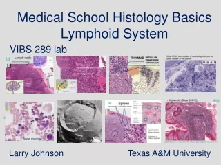
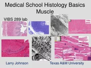
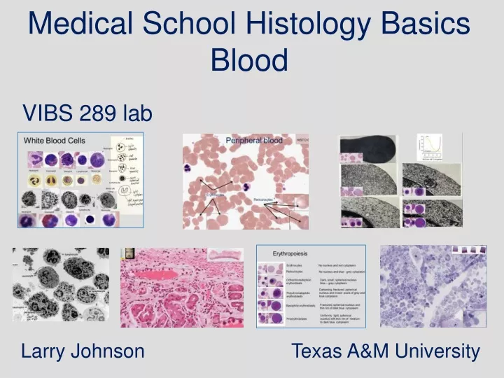
Medical School Histology Basics Blood VIBS 289 lab Larry Johnson Texas A&M University
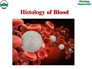
Blood (definition and function) Blood - fluid tissue composed of erythrocytes (RBC), leukocytes (WBC), and platelets suspended in blood plasma. Function of blood is the transportation of cells and fluid. RBC carry O2 to and CO2 from tissues.
Blood Function of blood is transportation of cells and fluid. WBC Immune defense for body. Plasma Nutrients to tissues. Waste from tissues. Proteins to hold water in plasma. Hormones and other informational mediators. Platelets Prevent loss of transportation.
Peripheral blood HISTO 021 Erythrocytes Reticulocytes
Reticulocyte Note ribosomes in the reticulocyte
Polychromatophilic normoblasts
Bone marrow 32583 Reticulocyte entering blood vessel Blood vessel
White Blood Cells Neutrophil Eosinophil Neutrophil Eosinophil Monocyte Basophil Basophil Lymphocyte Lymphocyte Monocyte Lymphocyte Monocyte Neutrophil Basophil Eosinophil
White blood cells in blood Eosinophil Basophil Neutrophil Lymphocyte Monocyte
Slide 113 Neutrophils platelets Neutrophils Red blood cells Large lymphocyte Lymphocyte Neutrophils Monocyte
SIZE OF HUMAN BLOOD CELLS CELL/PLATELETSIZE ERYTHROCYTES 6.5-8 µm LEUKOCYTES (WBC) % of WBC NEUTROPHIL 12-15 µm 60-70% LYMPHOCYTE 6-18 µm 25% MONOCYTE 12-20 µm 5% EOSINOPHIL 12-15 µm 2-4% BASOPHIL 12-15 µm 0-1% PLATELETS 2-4 µm
110 110 Peripheral blood smear (Leishman-Giemsa) Monocyte Neutrophils Monocyte Neutrophils Neutrophils Lymphocyte Monocyte
110 110 Peripheral blood smear (Leishman-Giemsa) basophil, eosinophil, and neutrophils Lymphocyte Neutrophils Basophil Neutrophils Neutrophils
110 Peripheral blood smear (Leishman-Giemsa)eosinophil and neutrophil platelets Red blood cells Eosinophil Neutrophil Eosinophil Neutrophil
113 Slide 113 Peripheral blood smear (May-Grunwald-Giemsa) Neutrophil Monocyte Lymphocytes Monocyte Lymphocyte
113 Peripheral blood smear (May-Grunwald-Giemsa) Monocyte Lymphocyte Monocyte Monocyte
Slide 113 Peripheral blood smear (May-Grunwald-Giemsa) Lymphocyte
HISTO 021 HISTO 021 60x
EM 8a EM 8c EM 8c illustrates an example of a basophil Note the neutrophils and eosinophils in EM 8a
140 Cardiac stomach w/chronic infection Infection site Plasma cells
140 140 Cardiac stomach w/chronic infection Neutrophil Eosinophil
EM 12b to observe organelle content differences in an immature and mature neutrophils – mature has more mature dumbbell-shaped granules EM 8f
EM 8f Lymphocytes
Note the relative paucity of cytoplasmic organelles and the high nuclear to cytoplasmic ratio (EM 6) EM 6 Lymphocytes
Large Golgi and many plasma membrane projects on monocytes. HISTO 021 Monocytes
Large Golgi and many plasma membrane projects on monocytes. 432 Macrophages (Slide 432) lung EM 8f Monocyte HISTO 021 1 Blood monocytes differentiate into macrophages like those seen in the lungs
Hematopoiesis Simplified: Part 1 Erythropoiesis https://www.youtube.com/watch?v=UTo4LnXonTk&list=PLQltfhuKi1sgtel3O9IpDsbgEWMY3JaX5&index=19 Larry Johnson Texas A&M University
Erythropoiesis Erythrocytes No nucleus and red cytoplasm Reticulocytes No nucleus and blue - grey cytoplasm Orthochromatophilic erythroblasts Dark, small, spherical nucleus blue – grey cytoplasm Darkening, fractured ,spherical nucleus and mixed pools of grey and blue cytoplasm Polychromatophilic erythroblasts Fractured, spherical nucleus and thin rim of dark blue cytoplasm Basophilic erythroblasts Uniformly light, spherical nucleus with thin rim of medium to dark blue cytoplasm Proerythroblasts
Erythropoiesis Proerythroblasts Erythrocytes Reticulocytes Orthochromatophilic erythroblasts Basophilic erythroblasts Polychromatophilic erythroblasts Erythropoiesis
Bone marrow 32583 Reticulocyte entering blood vessel Blood vessel
EM 12a Bone marrow
EM 8a marrow blood
432 Lung with bronchi – macrophage 432
432 Lung with bronchi 432