Download
1 / 48
480 likes | 494 Views
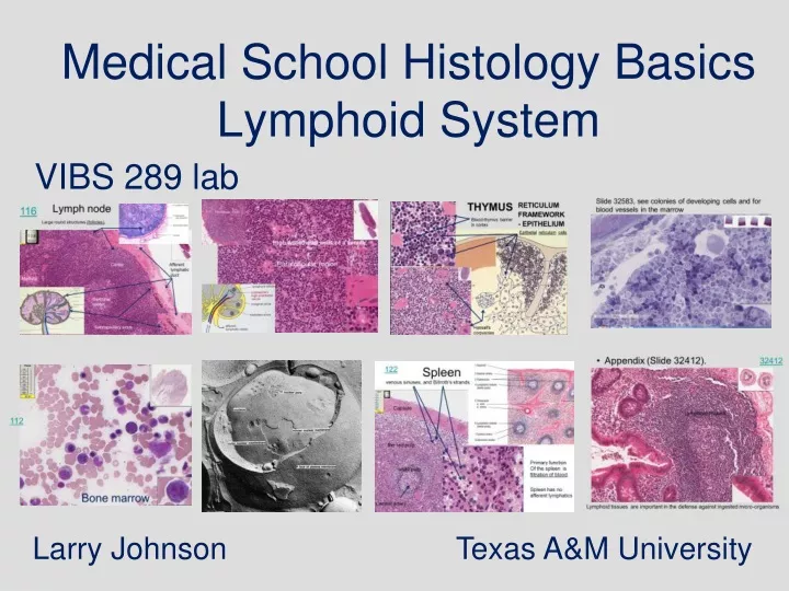
Medical School Histology Basics Lymphoid System. VIBS 289 lab. Larry Johnson Texas A&M University. EXAMPLES OF IMMUNE RESPONSE. REACTION AGAINST MICROORGANISMS: BACTERIA, VIRUSES, PARASITES REACTION AGAINST TUMOR CELLS

E N D
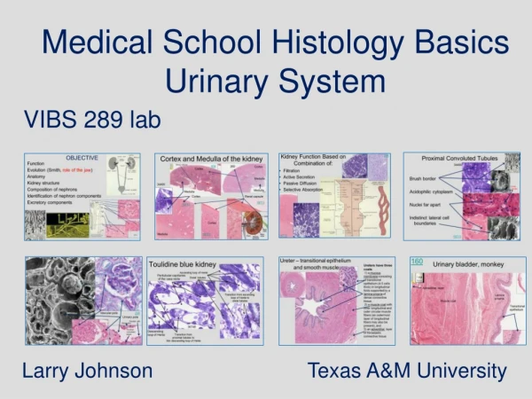
Medical School Histology Basics Lymphoid System VIBS 289 lab Larry Johnson Texas A&M University
EXAMPLES OF IMMUNE RESPONSE • REACTION AGAINST MICROORGANISMS: BACTERIA, VIRUSES, PARASITES • REACTION AGAINST TUMOR CELLS • ALLERGIC REACTIONS: HAY FEVER, POISON IVY • AUTOIMMUNE REACTION: ARTHRITIS, TYPE I DIABETES • GRAFT REJECTION Appendix 32412 http://www.greenlifestyle.be
PURPOSE OF THE IMMUNE SYSTEM CELLULAR BASIS OF IMMUNITY EFFECTORS OF RESPONSE INDUCTION OF THE RESPONSE ONTOGENY OBJECTIVES
FUNCTIONS OF THE IMMUNE SYSTEM • PROTECTION AGAINST FOREIGN INVADERS INTO BODY • PRODUCE / PROTECT GERM FREE ENVIRONMENT OF THE BODY
Three Key Steps of Combating Infections reak the cycle of transmission ill the infectious agent ncrease host resistance e.g., increase immunity of host
Lines of Defense First Line: Physical Barrier • Skin: Stratum Cornium • HCl In Stomach • Mucus In Intestines reak the cycle of transmission
Lines of Defense • Second line: Phagocytes at work • Neutrophils to ill the infectious agent Monocytes - macrophage
Characteristics of Immunity ncrease host resistancethrough Immunity Acquired - requires exposure to antigens Specificity - response is unique to exposure Memory - remembers previous exposure
Characteristics of Immunity Acquired - Must be developed Specificity - Antibodies made are specific to specific molecules on the antigen of exposure
Characteristics of Immunity Memory: quick second response • Long lived cells
Types of Immune Response • Antibody: mediated • Glycoproteins recognize and bind to antigens • Cell: mediated • Specifically active cells recognize cell - bind antigens
Slide 32583, see colonies of developing cells and for blood vessels in the bone marrow
112 Smear of bone marrow cells
thymus Bone marrow
123 Thymus, newborn Outer darkly staining areas (cortex) and lighter central areas (medulla). Medulla Thymocytes Cortex Medulla Continuous capillaries, sheathed by epithelial reticular cells around each, characterizes blood vessels in the thymus cortex and is responsible for the blood thymus barrier. Also there are no afferent lymphatics in the thymus.
123 Thymus, newborn epithelialreticulumcells Cortex Mitotic figures frequently, which reflect the high proliferative rate of these cells.
THYMUS Blood-thymus barrier in cortex Epithelialreticulumcells RETICULUM FRAMEWORK - EPITHELIUM Hassall's corpuscles
116 Lymph node Large round structures (follicles), Cortex Afferent lymphatic duct Medulla Germinal center, Capsule Capsule Subcapsulary sinus
Lymph node Capsule Afferent lymphatic duct 116 Capsule Subscapular space
Predominantly T lymphocytes Predominantly B lymphocytes, around germinal center Perifollicular area Germinal center High endothelial venules = sites where blood-borne lymphocytes enter the node.
Parafollicular region Parafollicular region Follicle 116 High endothelial venule Part of follicle Typical flat endothelium = Site of lymphocyte entry into the lymph node (one way street)
INDUCTION OF RESPONSE • PERIPHERAL ORGAN NEEDED TO GET ANTIGEN AND RESPONSIVE CELL TO INTERACT • LYMPHOCYTE RECIRCULATING • APPROPRIATE CONTEXT
19754 Vinous sinus in lymph node Vinous sinus Medullary cords
Parafollicular region Efferent lymphatic duct? 116
Tonsil 419
419 Tonsil
242 Esophagus and trachea, monkey
145 Fundic stomach
153 Plasma cells large intestine in Colon, monkey
32412 • Appendix (Slide 32412). Lymphoid tissues Lymphoid tissues are important in the defense against ingested micro-organisms
218 Spleen (reticulum stain)- capsule and reticulum fibers
122 Spleen Venous sinuses, and Billroth's strands Capsule the red pulp. Primary function Of the spleen is filtration of blood. white pulp Spleen has no afferent lymphatics Central artery
Penicillar arteries in marginal zone 117 Spleen Marginal zone Follicles White pulp Central artery Red pulp Venous sinuses Billroth's strands
Spleen 122 central artery PENICILLARY ARTERIES Marginal zone
218 Spleen (reticulum stain)- reticulum fibers in strands between venous (blood) sinus
Litteral cells of spleenic venule Spleen #19752 (UT117?) 117
Spleen Littoral cells: picket-fence type endothelial cells of vascular sinus Penicillar arteries Billroth’s strand or spleenic strand
Bone marrow 19761 Lymphocyte is a main player of immune response Blood in vessel
Life Cycle of Lymphocytes Fetal organs Bone marrow Primary lymphoid organs (Antigenindependent development) - Thymus – T lymphocytes - Bone marrow - B lymphocytes Secondary lymphoid organs (Antigendependent development) • Lymph nodes • Lymphoid nodules • Spleen
Questions on the lymphoid system 1. The main purpose(s) of lymphocyte traffic, the circulation of lymphocytes throughout the body, is: a. to place the responsible lymphocytes in the region of specific antigens b. to stimulate lymphocyte growth through meiosis c. to clean out the lymphatics ducts d. a and b e. a, b, and c 2. The function(s) of the spleen include: a. clean the blood of particulate matter b. to remove worn red blood cells c. to remove ribosomes from reticulocytes d. a and b e. a, b, and c 3. Which lymphatic organ(s) contain(s) penicilliary arteries or both afferent and efferent lymphatic vessels? a. lymph node b. spleen c. thymus d. a and b e. a, b, and c
Many illustrations in these VIBS Histology YouTube videos were modified from the following books and sources: Many thanks to original sources! • Bruce Alberts, et al. 1983. Molecular Biology of the Cell. Garland Publishing, Inc., New York, NY. • Bruce Alberts, et al. 1994. Molecular Biology of the Cell. Garland Publishing, Inc., New York, NY. • William J. Banks, 1981. Applied Veterinary Histology. Williams and Wilkins, Los Angeles, CA. • Hans Elias, et al. 1978. Histology and Human Microanatomy. John Wiley and Sons, New York, NY. • Don W. Fawcett. 1986. Bloom and Fawcett. A textbook of histology. W. B. Saunders Company, Philadelphia, PA. • Don W. Fawcett. 1994. Bloom and Fawcett. A textbook of histology. Chapman and Hall, New York, NY. • Arthur W. Ham and David H. Cormack. 1979. Histology. J. S. Lippincott Company, Philadelphia, PA. • Luis C. Junqueira, et al. 1983. Basic Histology. Lange Medical Publications, Los Altos, CA. • L. Carlos Junqueira, et al. 1995. Basic Histology. Appleton and Lange, Norwalk, CT. • L.L. Langley, et al. 1974. Dynamic Anatomy and Physiology. McGraw-Hill Book Company, New York, NY. • W.W. Tuttle and Byron A. Schottelius. 1969. Textbook of Physiology. The C. V. Mosby Company, St. Louis, MO. • Leon Weiss. 1977. Histology Cell and Tissue Biology. Elsevier Biomedical, New York, NY. • Leon Weiss and Roy O. Greep. 1977. Histology. McGraw-Hill Book Company, New York, NY. • Nature (http://www.nature.com), Vol. 414:88,2001. • Arthur C. Guyton,1971.Textbook of Medical Physiology W.B. Saunders company, Philadelphia, PA • WW Tuttle and BA Schottelius 1969 Textbook of Physiology C.V. Mosby Co. • A.L. Mescher 2013 Junqueira’s Basis Histology text and atlas, 13th ed. McGraw