Download
1 / 56
570 likes | 610 Views
Explore the anatomy of the kidney, nephron components, and the excretory function of the urinary system for homeostasis and waste removal. Learn about portal systems and the filtration barrier for urine formation.

E N D
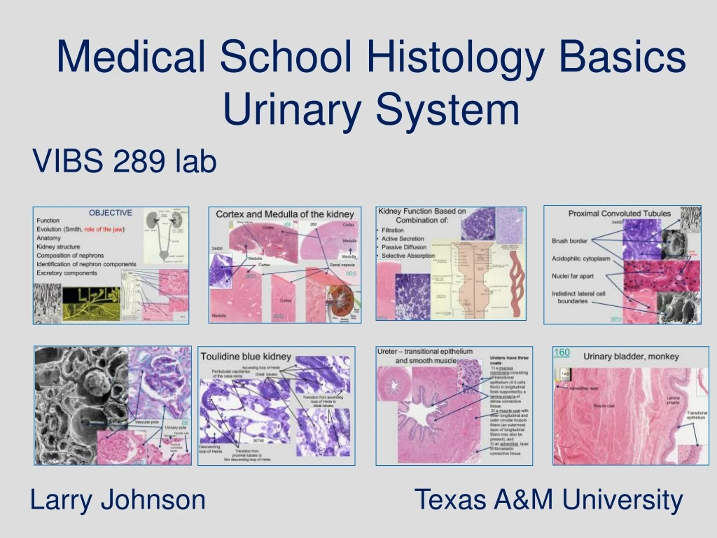
Medical School Histology Basics Urinary System VIBS 289 lab Larry Johnson Texas A&M University
OBJECTIVE Function Evolution (Smith, role of the jaw) Anatomy Kidney structure Composition of nephrons Identification of nephron components Excretory components
Function of Urinary System: Homeostasis Rid body of waste (urea, uric acid, creatinine, salts) Preserves constancy of extracellular fluid in composition, volume, and pH Endocrine function • Secrete erythropoietin - red blood cell production • Produces renin - aldosterone release
258 Kidney Function Based on Combination of: 34400 • Filtration • Active Secretion • Passive Diffusion • Selective Absorption 19713
Evolution – Animals escaping predators Fresh water Salt water
PORTAL ARTERIOLE
ARTERIAL PORTAL SYSTEM Portal system = CAPILLARY PORTAL ARTERIOLE CAPILLARY afferent ARTERIOLE Glomerular CAPILLARY efferent ARTERIOLE and PERITUBULAR CAPILLARIES Function of a portal system? local change in blood composition whereby the first capillary modifies and second allows the change in composition to affect local cells near it. 1st 2nd 1st 2nd
Cortex Medulla Cortex 19713
Hilus region where blood vessels enter and leave and the ureter begins
Cortex and Medulla of the kidney Minor calyces 458 260 Cortex Cortex Medulla 34400 Medulla Medulla Cortex Renal capsule 19713 19713 Cortex Medulla 19713
Cortex Medulla 258
Cortex Medulla 258
19713 kidney thin loop of Henle in cortex Thin loop of Henle in cortex
Nephrons are the structural and functional units of the kidney. Portal Nephrons consists of: • A glomerulus, • Bowman's capsule, • proximal convoluted tubule, • loop of Henle, • distal convoluted tubule, and • collecting tubule (collecting ducts).
Nephrons are the structural and functional units of the kidney. 34400
Cortex 19713 Medulla
Slide Histo 032: Kidney (H&E) Urinary pole Bowman's space Parietal epithelium Bowman’s capsule Podocyte Glomerulus Renal corpuscle Visceral epithelium Arteriole of vascular pole Juxtaglomerular cells Macula densa
Renal corpuscle The renal corpuscle provides the anatomical structure required for the first phase of urine formation: the production of the glomerular filtrate. Several histological arrangements and structures are required for the production of glomerular filtrate: 1) arterioles entering and leaving the glomerulus, 2) glomerular endothelium, 3) glomerular basal lamina, and 4) visceral epithelium of the Bowman's capsule Arterioles 19713 Visceral epithelium Distal tubule and macula densa Distal tubule and macula densa Glomerular endothelium Bowman's space Glomerular basal lamina 258
Components of the filtration barrier: 1. The fenestrations of the capillary endothelium, which blocks blood cells and platelets 2. The thick, combined basal laminae, or GBM, which restricts large proteins and some organic anions 3. The filtration slit diaphragms between pedicels, which restrict some small proteins and organic anions The glomerular basal lamina, thick and anionic, filters macromolecules according to their size and electrostatic charge. EM 18a
Glomerular Features for Extreme Filtration 258 Very Large Surface Area (1.5 m2) Large resistance afforded by reduced diameter of the efferent arteriole Thin Filter (0.1 µm) Thus, 25 times more permeable than regular capillaries 19713 Glomerular basal lamina The hydrostatic pressure within the glomerular capillaries provides the driving force for producing 180 liters of glomerular filtrate per day. The filtrate resembles blood plasma without large (>40,000 MW) proteins. Endothelium of the glomerular capillaries is fenestrated
Urinary pole – site of union of parietal cells of Bowman's Capsule and cells lining the proximal convoluted tubules Vascular pole – site of afferent and efferent arterioles
Vascular pole 258 Urinary pole Parietal cells Glomerulus Proximal convoluted tubule 19713 EM 18b
Slide Histo 032: Kidney (H&E) Juxtaglomerular apparatus Macula densa of distal tubule Juxtaglomerular cells Distal convoluted tubules Proximal convoluted tubules Microvilli Brush border
Brush border is involved in reabsorption of protein. Proximal convoluted tubules 19713 0 to 8 mg/dL protein in urine is considered normal. 258 Brush border
Proximal Convoluted Tubules 34400 Brush border Acidophilic cytoplasm Nuclei far apart Indistinct lateral cell boundaries Distal tubules Histo32 19713
Kidney (H&E) Cortex Capsule Cortex Medulla Medulla Arcuatearteries Juxtamedullary nephrons Medullary rays Slide Histo32
Transitions 36748
Toulidine blue kidney Ascending loop of Henle Peritubular capillaries of the vasa recta Distal tubules Transition from ascending loop of Henle to distal tubules 36748 Descending loop of Henle Transition from proximal tubules to thin descending loop of Henle
36748 Transitions from ascending loop of Henle to distal tubules Enfolding of the basal cell membrane line up mitochondria in cells of the distal tubule.
Distal Convoluted Tubules No brush border Less acidophilic cytoplasm Nuclei relatively close Larger lumen
258 Renin granules in JG cells of Kidney (PAS)
Rich blood supply of peritubular capillaries and vasa recta cortex 260 Arcuate arteries medulla
36748 Ascending loop of Henle Distal tubule Blood capillaries of the vasa recta Descending loop of Henle
Countercurrent exchanger Capillaries of the vasa recta
Peritubular Capillaries Absorbs - 180 liters/day from interstitial spaces; thus, ~4 times reabsorption of venous end of all other capillaries of body Endothelial cells - extremely porous Colloidal osmotic pressure of plasma proteins Low capillary pressure Proximity to uriniferous tubules Peritubular Capillaries About 85% of the water and sodium of the glomerular filtrate is resorbed by the proximal convoluted tubule and passes back into the bloodstream via the peritubular capillaries.
Kidney medulla CT CT TnLH ThLH CT CT Cap CT Cap 19713
458 kidney Collecting duct
Slide Histo 032: Kidney (H&E) Apex of renal pyramid Renal calyx CT adventia capsule Calyx Renal pyramind and calyx Medulla Renal pelvis Cortex Histo32 Simple columnar to transitional-like epithelium of pyramid
458 kidney cortex medulla Minor calyx
Ureter – transitional epithelium and smooth muscle Ureters have three coats: 1) a mucous membrane consisting of transitional epithelium (4-5 cells thick) in longitudinal folds supported by a lamina propria of dense connective tissue, 2) a muscle coat with inner longitudinal and outer circular muscle fibers (an outermost layer of longitudinal fibers may also be present), and 3) an adventitial layer of fibroelastic connective tissue
160 Urinary bladder, monkey Adventitial layer Lamina propria Muscle coat Transitional epithelium
277 Human Penis – transitional epithelium and surrounding spongy cavernous of penal urethra