Download
1 / 21
220 likes | 428 Views
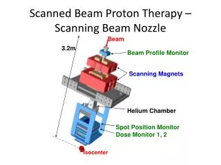
Optimisation in proton scanning beams. First results…. Basic steps. Calculation of the dose-deposition coefficients (ddc’s) Optimisation of the spot weights. Spots and beamlets. Beam beamlets or pencil beams (defined by the resolution of the calculation grid)

E N D
Optimisation in proton scanning beams First results…
Basic steps • Calculation of the dose-deposition coefficients (ddc’s) • Optimisation of the spot weights
Spots and beamlets • Beam beamlets or pencil beams (defined by the resolution of the calculation grid) • The dose from each beamlet is evaluated (at the vertices of the calculation grid) • The spot dose is calculated (as the sum of the dose contributions of the corresponding beamlets, weighted for the position of each beamlet within the spot)
Sx and Sy depend on z spot depends on the density and on z beamlet
Optimisation: a closer look • Desired dose at point i: pi • Dose delivered at point i: di = aij xj (sum over all sources j) Objective function: Fobj = (di - pi)2 (sum over target points) + contribution due to the violation of dose-limit constraints (for targets and organs) + contribution due to the violation of dose-volume constraints (for organs)
Optimisation methods • Conjugate Gradient (CG) • Simulated Annealing (SA)* • ‘Simultaneous’ optimisation (PSI) • Generalised Sampled Pattern Matching (GSPM)* (* = under development)
Strategy in the optimisation • Pre-optimisation Reasonable initial ‘guess’ for the weights Convergence two consecutive iterations yield improvement below 5% • Main optimisation Full implementation of a method Convergence two consecutive iterations yield improvement below 0.1%
Toy example • A phantom has been created with three important structures: one target and two organs; some inhomogeneity has been introduced (an additional structure simulating the presence of a bone) • Pixel size: 2.5mm • Spot advance in y (scanning direction): 2.5mm • Spot advance in x: 5mm • Cut-off for dose contributions: 3 standard deviations
Target: 2,412 points, 57.27 cm3 Distal Organ: 2,166 points, 48.34 cm3 Proximal Organ: 683 points, 15.49 cm3 Number of points: 5,261
Dose-Volume Histograms Prescription dose: 50 Gy ( 2%) Organ constraints: 25 Gy in 10% of the distal organ;15 Gy in the proximal organ
Weight distribution PSI method
Weight distribution SA method
Conclusions • As far as the dose distribution is concerned, three optimisation methods (CG, SA, and PSI) yield results which seem to be in good agreement. Very similar dose distributions may be obtained on the basis of very different weight distributions. • The use of raw (unfiltered) weights does not seem to create cold/hot spots within the irradiated volume. It remains to be seen whether, in some occasions, filtering will be called for.
Under consideration… • Other forms of the objective function to be tried? • Strategy in the optimisation: an improvement of about 25% was found in the execution time in case that the target dose is firstly optimised (with vanishing dose everywhere else) • Other optimisation methods to be tried?