Download
1 / 84
970 likes | 1.87k Views
Myology. SHANDONG UNIVERSITY Liu Zhiyu. Myology. Morphology of skeletal muscle Muscle belly Tendon aponeurosis Classification Long muscle Short muscle Broad muscle Orbicular muscle. Myology. Origin - the fixed attachment Insertion - the movable attachment Action

E N D
Myology SHANDONG UNIVERSITY Liu Zhiyu
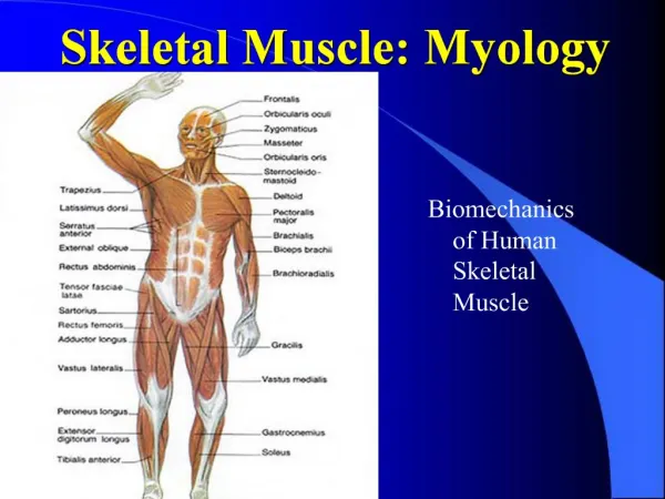
Myology • Morphology of skeletal muscle • Muscle belly • Tendon aponeurosis • Classification • Long muscle • Short muscle • Broad muscle • Orbicular muscle
Myology • Origin -the fixed attachment • Insertion -the movable attachment • Action • Agonist • Antagonist • Synergist • Fixators • Nomenclature of mucles : • shape • size • Location • their points of attachment
Myology Accessory structures • Fascia • Superficial fascia • Deep fascia • Synovial bursa
Myology • Tendinous sheath • Fibrous layer • Synovial layer: • Mesotendon • vincula tendinum
Facial muscles • Epicranius • Frontal belly • Occipital belly • Galea aponeurotica • Orbicularis oculi • Buccinator • Orbicularis oris • Nasalis
★Masticatory muscles • Temporalis • Masseter • lateral pterygoid • Medial pterygoid
★ Masticatory muscles • Temporalis • Origin-temporal fossa • Insertion-coronoid process of mandible • Action-elevates and retracts mandible • Masseter • Origin-inferior border and medial surface of zygomatic arch • Insertion-lateral surface of ramus of mandible and angle of mandible • Action-elevates mandible
★ Masticatory muscles • Lateral pterygoid • Medial pterygoid
Muscles of the neck Superficial group • Platysma 颈阔肌 • Sternocleidomastoid 胸锁乳突肌
Muscles of the neck Suprahyoid muscles • Digastric • Mylohyoid • Stylohyoid • Geniohyoid Elevate (raise) hyoid bone and depress mandible.
Muscles of the neck Infrahyoid muscle • Sternohyoid • Sternothyroid • Thyrohyoid • Omohyoid Depress hyoid or larynx after elevation
Muscles of the neck Deep group • Lateral • Scalenus anterior • Scalenus medius • Scalenus posterior • Medial • longus capitis • longus colli Flex the head, bends the neck forward
Major muscles of the neck ★ Sternocleidomastoid • Origin: manubrium and sternal end of clavicle • Insertion: mastoid process of temporal bone • Action: contraction of one muscle draws head toward the same side, and turn face to opposite side; both muscles act together to draw head backward
Major muscles of the neck Scalenus anterior • Origin: transverse processes of C3-C6. • Insertion: tubercle for scalenus anterior • Action: unilateral, bends neck laterally; bilateral, elevate first rib, an accessory muscle of inspiration; if rib is fixed, flex neck anteriorly
Major muscles of the neck ★ Scalene fissure • Above the first rib, there is a triangular space between scalenus anterior and medius. • The brachial plexus and the subclavine a. emerge from this space.
The Muscles of Back Superficial group • Trapezius • Levator scapulae • Rhomboideus • Latissimus dorsi • Thoracolumbar fascia
The Muscles of Back Deep group • Splenius • Erector spinae
Major Muscles of Back Trapezius • Origin: superior nuchal line, external occipital protuberance, ligamentum nuchae and spinous processes of seventh cervical and all thoracic vertebrae • Insertion: lateral third of clavicle, acromion, and spine of scapular • Acton: upper fibers elevate scapula, lower fibers depress scapula; if scapula is fixed, one side acting along, draws head toward the same side, and turn face to opposite side; both sides together, draw head directly backward • Nerve supply: accessory nerve (Ⅺ cranial nerve)
Major Muscles of Back Latissimus dorsi • Origin: • Spinous processes of lower six thoracic and all lumbar vertebrae • Median sacral crest • Posterior part of iliac crest • Insertion: floor of intertubercular groove of humerus. • Action: trunk fixed, extends, adducts and medially rotates arm ; arm fixed, elevates trunk. • Nerve supply: thoracodorsal nerve
Thoracolumbar Fascia Anterior layer Middle layer Posterior layer
Trapezius Levator scapular Deltoid Ausculatory triangle Rhomboideus Latissimus dorsi Thoracolumbar fascia Inferior lumbar triangle
Muscles of thorax Muscles connecting the upper limb to the thoracic wall • Pectoralis major • Pectoralis minor • Serratus anterior
Muscles of thorax • Intrinsic muscles • Intercostales externi • Intercostales interni • Intercostales intimi • Transverses thoracis
Majormuscles of thorax Pectoralis Major • Origin: medial half of clavicle, sternum, upper six costal cartilages. • Insertion: lateral lip of the bicipital groove of humerus • Action: adducts the arm and rotates it medially; the clavicular fibers also flex the arm; with the arm above the head, raise the body as in climbing • Nerve supply: lateral pectoral n.
Majormuscles of thorax Intercostales externi • Origin: inferior border of rib above • Insertion: superior border of rib below • Replaced anteriorly by external intercostals membrane • Action: raise ribs adding in forced inspiration
Major muscles of thorax Intercostales interni • Origin: superior border of rib below • Insertion: inferior border of rib above • Replaced posteriorly by internal intercostals membrane. • Action: depress ribs for forced expiration
Diaphragm • Shape and position: dome-shaped between thorax and abdomen, consists of a peripheral muscular part and a central tendon • Origin • Sternal part: arising from xiphoid process • Costal part: arising from lower six and costal cartilages • Lumbar part: arising by two crura from upper 2-3 lumbar vertebrae • Insertion: central tendon • Weak areas: • Lumbocostal triangle • Sternocostal triangle
Diaphragm Openings in the diaphragm • Aortic hiatus lies anterior to the body of the 12th thoracic vertebra between the crura and transmits the aorta, thoracic duct • Esophageal hiatuslies at level of T10. It transmits esophagus and vagus nerves • Vena cava foramenlies at T8 level in the central tendon. It transmits the inferior vena cava. T8 T10 T12
Diaphragm Action: • Contraction: the dome moving downward, increases the volume of thoracic cavity which results in inspiration, at the same time the intra-abdominal pressure is increased assists in defecation, vomiting or child birth. • Relaxation: the dome returns to the former position, reduces the volume to the thoracic cavity, resulting in expiration.
Muscles of abdomen Anterolateral group • Obliquus externus abdominis • Obliquus internus abdominis • Transversus abdominis • Rectus abdominis
Obliquus externus absominis • General direction of fibers: downward, forward and medially (run down and inward)
Obliquus externus absominis Structures • Inguinal ligament • Lacunar ligament • Superficial inguinal ring-triangular-shaped defect in aponeurosis of obliquus externus abdominis above pubic tubercle
Obliquus internus abdominis • Deep to obliquus externus abdominis • General direction of fibres: upwards, forwards and medially
Transversus abdominis • Deep to obliquus internus • General direction of fibers: run horizontally forward.
Transversus abdominis • Inguinal falx • Obliquus internus abdominis has a lower, free border that arches over spermatic cord • Inserted with transversus abdominis fiber into medial part of pecten of pubis • Cremaster • Dirived from the lower fibers of the obliquus internus abdominis and transversus abdominis • Around the spermatic cord and testis
Rectus abdominis • Position: lie on to either of midline • Origin: pubic crest and symphysis • Insertion: xiphoid and 5th-7th costal cartilages • Tendinous intersections3-4 • linea semiluaris
Similar functions for above four pairs of muscles • Support and compress the abdominal viscera • Increase intra-abdominal pressure, aid in expulsive efforts-vomiting, coughing, sneezing, defecation, urination and childbirth. • Depress ribs, assist in (the act of force(4)expiration. • Flex, lateral flex, and rotate vertebral column
Sheath of rectus abdominis Anterior layer • Formed by fusion of aponeurosis of obliquus externus abdominis and anterior leaf of aponeurosis of obliquus internus abdominis
Sheath of rectus abdominis Posterior layer • Formed by fusion of posterion leaf of aponeurosis of obliquus internus abdominis and aponeurosis of transversus abdominis • Absent in about 4-5cm below the umbilicus, where aponeuroses of all three muscles form anterior layer the lower free border named arcuate line • Below this line rectus abdominis in contact with transverse fascia
Muscles of abdomen • Linea alba -tendinous raphe between right and left recti from xiphoid to pubic symphysis
Landmarks and surface anatomy • Linea alba • Rectus abdominis • Tendinous intersections • Linea semilunaris • Umbilicus: at the level of L3 ~ L4 • Inguinal ligament
Muscles of abdomen Posterior group • Quadratus lumborum • Psoas major