Download
1 / 26
260 likes | 895 Views
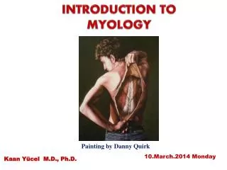
PRINICIPLES OF MYOLOGY. Def: science deals with muscles. Skeletal muscles are the active part of locomotor system Smooth muscles move the viscera Cardiac muscle produces heart contraction. Dr: Ahmed Saber Vet. Anat. 1. 2008. The main characters of muscles are:

E N D
PRINICIPLES OF MYOLOGY Def: science deals with muscles • Skeletal muscles are the active part of locomotor system • Smooth muscles move the viscera • Cardiac muscle produces heart contraction Dr: Ahmed Saber Vet. Anat. 1. 2008
The main characters of muscles are: Irritability or Excitability - responsive to chemical, electrical, or mechanical stimuli Contractility - ability of muscle to contract (shorten) Extensibility - ability to be passively stretched Elasticity - ability to return to its original length Dr: Ahmed Saber Vet. Anat. 1. 2008
Types of muscles 1- smooth muscles: • Structures: long spindle shaped, uni-nucleate, and non-striated (smooth) • Locations: wall of visceral organ • The control:involuntary autonomic control or endocrine control (myo-epithelial cells) • function: Involuntary contractions that move materials through the organs Dr: Ahmed Saber Vet. Anat. 1. 2008
2- Cardiac muscles • Structures: • cylindrical, mostly uninucleate striated, and Branching and joins forming myocardium network (syncytium). • The fibers are connected end-to-end at sites called intercalated disks. Dr: Ahmed Saber Vet. Anat. 1. 2008
Locations: wall of heart • The control:involuntary • function: Involuntary autonomic control and special local conducting system cause contractions of heart Dr: Ahmed Saber Vet. Anat. 1. 2008
3- Skeletal muscles They forming about 40-45 % of body weight The muscle cell = muscle fiber = myofiber • structures: • Elongated, multinucleated, with long and transverse striation. • Each muscle is divided into bundles or fascicles • Each fascicles is composed of numerous muscle fiber (cells) • Locations: in all body & forming active part of locomotor system Dr: Ahmed Saber Vet. Anat. 1. 2008
Skeletal muscles (cont.) • The control:voluntary control function: • voluntary somatic movement of the body • Maintain body position • Stabilizing the joint • Heat production • Guarded body entrances and orifices Dr: Ahmed Saber Vet. Anat. 1. 2008
Fiber Fascicle Epimysium Muscle Tendon bone Architecture of skeletal muscle • Each single muscle is consists of numerous bundles (fascicles) • Each fascicles is consists of numerous muscle fibers • The muscle fiber is the structural unite of muscle Dr: Ahmed Saber Vet. Anat. 1. 2008
Architecture of skeletal muscle Muscle fascicle Blood vessels Epimysium Muscle fiber &endomysium Perimysium Epimysium: CT layer ensheated the entire muscle Perimysium: CT layer ensheated each muscle bundle (fascicle) Endomysium: CT layer ensheated each muscle fiber Epimysium, perimysium and endomysium extend at the muscle end forming tendon or aponeurosis They protect the cells and provide passage for vessels and nerves Dr: Ahmed Saber Vet. Anat. 1. 2008
Architecture of skeletal muscle Muscle fiber Perimysium Fascicle Endomysium Epimysium Dr: Ahmed Saber Vet. Anat. 1. 2008
Microscopic structures Myofibril Myofiber Myofilaments Actin Myosin Dr: Ahmed Saber Vet. Anat. 1. 2008
Gross anatomy of skeletal muscle 1- anatomical parts: A- origin: • usually the proximal attachment of muscle • Less movable part • Somtime more that one head (biceps) B- insertion: • Usually distal attachment • More movable C- belly: • The main balk of muscle between origin and insertion Dr: Ahmed Saber Vet. Anat. 1. 2008
Gross anatomy of skeletal muscle 2- skeletal muscle attachment • Bone • Cartilage • Deep fascia (indirectly to bone) • Skin and superficial fascia • Intermediate tendon • No attachment (no origin & no insertion, as orbicularis oris) Also muscle attachment may be Direct attachment: the epimysium is fused to periosteum or perichondrium directly Indirect attachment: epimysium extends as sheet like aponeurosis before attaching to bone, cartilage, fascia or other muscle Dr: Ahmed Saber Vet. Anat. 1. 2008
Gross anatomy of skeletal muscle 3- arrangements of skeletal muscle fibers Based on the angle between fiber and the line of pull • A- Parallel fibers: • Strap (sartorius) • Quadrates (quadratus femoris) • Fusiform (biceps brachi Strap Fusiform Dr: Ahmed Saber Vet. Anat. 1. 2008
Unipennate B- oblique fibers (pennation) The fibers attached to tendon at oblique angle (feather like) 1- unipennate: on one side of tendonex extensor digitorun longus M 2- bipennate : on opposite sides of tendon, ex rectus femoris M 3- multipennate: muscle fibers attached to several fibrous bands within the muscle, the bands join forming one tendon, ex deltoid M 4- Centripennate: one central tendon Bipennate Multipennate Dr: Ahmed Saber Vet. Anat. 1. 2008
C- circular fibers 1- circular: no attachment, around orifices, ex orbicularis oris (around mouth) 2- convergent: broad origin and pointed insertion, ex pectoralis major Circular Convergent
Functional types of muscles • Prime mover (Agonist muscles) • muscles most involved • cause joint motion through a specified plane of motion when contracting • Antagonist muscles • located on opposite side of joint • have the opposite action to agonist Dr: Ahmed Saber Vet. Anat. 1. 2008
3. Synergist • assist in action of agonists • not necessarily prime movers for the action 4. Fixators (stabilozers) • Fix the joint Dr: Ahmed Saber Vet. Anat. 1. 2008
Blood and nerve supply • Vessels and nerves pass through the connective tissue sheaths • Bell is more vascular • One neuron supply variable number of muscle fibers • Motor unites = neuron+ muscle fiber innervated by this nerun Dr: Ahmed Saber Vet. Anat. 1. 2008
Accessory structures of skeletal muscle 1. Sesamoid bone • In course of some tendon • as superficial and deep flexor tendons • Protect the tendon • Decrease friction • Redirect the pull angle of the tendon Dr: Ahmed Saber Vet. Anat. 1. 2008
Body wall Internal fascia Bodycavity Deep fascia skin Superficial fascia 2. Fascia • is sheath of CT found all over the body • Divided into • Internal fascia (endothoracic, abdominaland pelvic) • External fascia ( superficial and deep fascia) Dr: Ahmed Saber Vet. Anat. 1. 2008
Synovial sac Tendon Bone 3. Synovial sac Is sac filled with synovia Located between two structures to decrease the friction between these structures • Subtendinous • Subcutaneous • subligamentous Dr: Ahmed Saber Vet. Anat. 1. 2008
4. Tendon synovial sheath • elongated sac under the tendon • The edges of the sac become reflected around the tendon • Consists of • Outer fibrous layer • Double inner synovial • Parietal and visceral layer in between small cavity filled with synovia • At edges meeting, the mesotendon present Dr: Ahmed Saber Vet. Anat. 1. 2008
Mesotendon Outer fibrous Parietal synoviallayer tendon Visceral synoviallayer Synovial fluid 4. Tendon synovial sheath Dr: Ahmed Saber Vet. Anat. 1. 2008
5. Retinaculum • Transverse band of deep fascia around carpal and tarsal joints • It fix the tendon and ligaments in its position • As, flexor and extensor retinaculum of carpus Dr: Ahmed Saber Vet. Anat. 1. 2008