Download
1 / 13
140 likes | 175 Views
Learn how muscles are named based on their location, size, shape, and action. Discover the naming conventions for muscles in the head and their innervation. Explore key terms like rectus, oblique, trapezoid, and more.

E N D
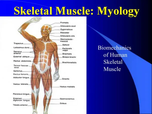
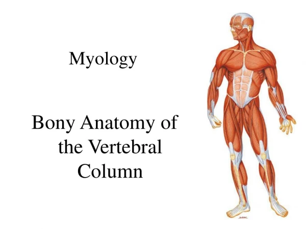
Myology Muscles of the Head
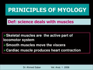
NAMING OF MUSCLES 1. Direction of muscle fibers: a. Rectus: fibers run parallel to the midline (rectus abdominis) b. Transverse: fibers run perpendicular to the midline (transverse abdominis) c. Oblique: fibers run diagonally to the midline (external oblique) 2. Location: a. named for a nearby bone: (frontalis) frontal bone b. position relative to a bone: (tibialis anterior) tibia c. section of a muscle having more than one part (anterior deltoid)
NAMING OF MUSCLES (cont) 3. Size: a. large muscle (gluteus maximus) b. small muscle (gluteus minimus) c. long muscle (extensor digitorum longus) d. short muscle (peroneus brevis) 4. Number of origins: a. two origins: biceps femoris b. three origins: triceps brachii c. four origins: pronator quadratus
NAMING OF MUSCLES(cont) 5. Shape: a. triangular: deltoid b. trapezoid: trapezius c. saw-toothed: serratus d. rhomboid or diamond shaped: rhomboids 6. Origin and Insertion: a. sternum, clavicle, and mastoid process: SCM
NAMING OF MUSCLES (cont) 7. Action: a. decreases an angle of a joint: flexor digitorum b. increases an angle of a joint: extensor carpi ulnaris c. moves bone away from midline: abductor pollicis brevis d. moves bone closer to midline: adductor longus e. upward movement: levator scapulae f. downward movement: depressor labii g. turns palm up or anteriorly: supinator h. turns palm down or posteriorly: pronator i. decreases size of an opening: external anal sphincter j. makes a body part more rigid: tensor fascia latae k. moves a bone around its longitudinal axis: rotator
Divided into 3 groups Muscles of the Scalp Muscles of the Face (Eyes, Nose, and Mouth) Muscles of Mastication Muscles of Head Overview
Muscles of the Head Overview • Muscle of head are located superficially in the fascia and therefore move the fascia of the scalp and/or face. • Muscles of scalp move scalp, ears, eyebrows • Muscles of facial expression subdivided into muscles that move: • Skin around eyes • Skin around nose • Skin around mouth • Muscles of mastication attach to mandible which is necessary for chewing i.e. mastication.
Muscles of Head Overview • Innervation: • Muscles of scalp innervated by facial nerve (CNVII). • Muscles of facial expression innervated by facial nerve (CN VII) • Muscles of mastication innervated by trigeminal nerve (CN V).
Muscles of the Scalp Occipitofrontalis: Can be considered 2 separate muscles the occipitalis and frontalis Both attach into the galea aponeurotica Galea aponeurotica is broad flat tendon also known as the epicranial aponeurosis Temporoparietalis: Also attaches into galea aponeurotica. Degree of development varies; some people it is thin other it is non- existent. Occipitofrontalis and temporoparietalis are known as the epicranius Auriculares: One, two, or all three are nonfunctional in many people Smallest is anterior auricularis, largest is posterior auricularis
Occipitalis O: Occipital & temporal bones I: Galea aponeurotica A: draws scalp posteriorly I: CN VII (Facial nerve) Palpation: page 15
Frontalis O: Galea aponeurotica I: Fascia and skin superior to the eye and nose A: draws scalp anteriorly and elevates the eyebrows I: CN VII (Facial nerve) Palpation: page 15
Temporoparietalis O: Fascia superior to ear I: Lateral border of galea aponeurotica A: Elevates ear and Tightens the scalp N: CN VII (Facial nerve) Palpation: page 18
Auricularis O: Anterior: anterior fascia of the ear Superior: lateral margin of the galea aponeurotica Posterior: temporal bone I: Anterior: Spine of helix Superior: Superior aspect of cranial surface of the ear Posterior: Posterior ear A: Anterior: draws ear anteriorly Superior: elevates ear Posterior: draws ear posteriorly N: CN VII (Facial nerve) Palpation: page 20