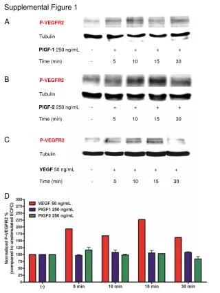
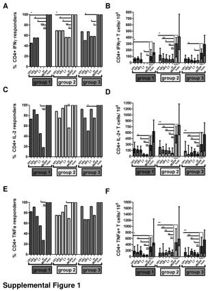
Download
1 / 1
10 likes | 107 Views
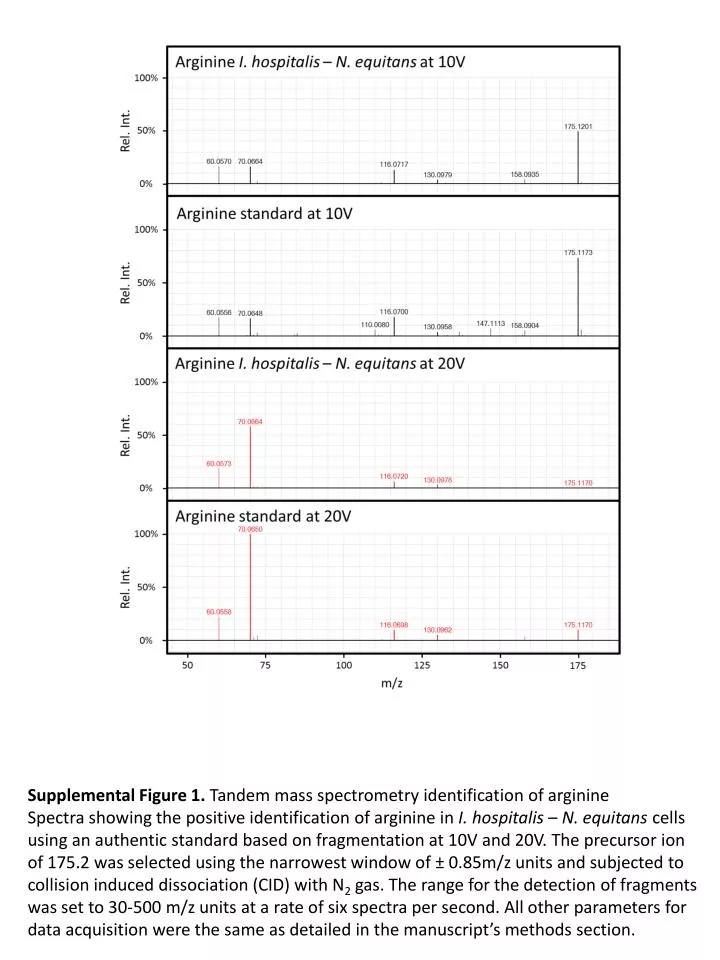
Supplemental Figure 1. Tandem m ass s pectrometry identification of arginine

E N D
Supplemental Figure 1. Tandem mass spectrometry identification of arginine Spectra showing the positive identification of arginine in I. hospitalis– N. equitanscells using an authentic standard based on fragmentation at 10V and 20V. The precursor ion of 175.2 was selected using the narrowest window of ± 0.85m/z units and subjected to collision induced dissociation (CID) with N2 gas. The range for the detection of fragments was set to 30-500 m/z units at a rate of six spectra per second. All other parameters for data acquisition were the same as detailed in the manuscript’s methods section.