Download
1 / 69
690 likes | 705 Views
Explore the intricate details and functions of the male and female reproductive systems, from spermatogenesis to oogenesis, and hormonal control to menstrual cycles. Learn about the production of sperm, ova, and the journey towards fertilization.

E N D
Main functions: The production of sperm, the male gametes; Spermatogenesis begins at puberty and continues until death; Delivery of functional sperm to the female reproductive system Anatomy of the human male
Anatomy of the human male Production of Semen (a fluid comprised of): Sperm, which are expelled through the ducts during ejaculation (≈1% of semen volume); Glandular secretions that carry, nourish, and protect the sperm (mostly sugars & buffers) Testes (plural): Glands that produce sperm; located outside abdominal cavity within the scrotum • Scrotum ≡ (saclike pouch ≈ 1-3°C below normal body temperature - sperm can only form at this lower temperature)
How sperm leave the testes Seminiferous tubules: carries/stores sperm in testes Epididymis: a series of coiled ducts for maturation & temporary storage of sperm Vas deferens: tube which carries sperm past connecting lubricating and support glands
How sperm leave the testes Lubricating and support glands: (a.) Seminal vesicles: secrete sugar-rich fluid that protects & nourishes sperm (b.) Prostate gland: produces an alkaline fluid that neutralizes both urine in the male urethra and the acidic environment of the vagina (c.) Bulbourethral glands: secrete fluids that lubricates the male urethra and allows easier coitus (intercourse)
How sperm leave the testes Urethra: tube in the penis that transports semen out of the male’s body; also transports urine from the urinary bladder Penis: copulatory organ; transient tumescence Ejaculation: the release of semen through rhythmic contractions of smooth muscle in the Vas deferens
Main functions: To produce the female gametes (ova); To receive sperm; To provide a suitable environment in which a fertilized ovum might developduring pregnancy Anatomy of the human female
Ovaries: Contain follicles that nurture ova; Produces sex hormones; Functional from puberty to menopause Anatomy of the human female
Oviducts (Fallopian Tubes): Convey (move) ova towards the uterus; Muscular contractions & cilia draw ovum (egg) into oviduct; Location for fertilization to occur Anatomy of the human female
Uterus (womb): Nourishes development of fertilized zygote; Opens into the vagina at cervix Vagina: Receives the penis during coitus; Forms the birth canal; Multiple layers of expandable smooth muscle; Potential, not defined, space Anatomy of the human female
Puberty Puberty: when secondary sexual characteristics develop and the potential for sexual reproduction is reached (sperm production or ovulation) Changes are controlled by hormones that initiate development of secondary sex characteristics; Primary sex characteristics are internal and external reproductive organs (genitalia)
Puberty in males (♂) Secondary sex characteristics: Primary hormone: testosterone (testes); 2° Characteristics: increased hair (body, pubic, & facial), muscle development, deeper voice; Spontaneous ejaculation
Puberty in females (♀) Secondary sex characteristics: Primary hormone: estrogen (ovaries); 2° Characteristics: breast development, broadened pelvis, distribution of body fat; increased hair (body & pubic); Menarche (onset of menstruation)
Reproductive hormones Testosterone (♂ testes): Sperm production & secondary sexual characteristics Estrogen (♀ ovaries); Ova production, preparing uterus for fertilized zygote & secondary sexual characteristics
Androgens, (testosterone most important), stimulate sperm production They also maintain homeostasis by a negative feedback mechanism that inhibits the secretion of FSH (follicle-stimulating hormone) and LH (luteinizing hormone) Hormonal control of the testes Stimuli from otherareas in the brain Hypothalamus Releasinghormone Anteriorpituitary Negative feedback FSH LH Androgenproduction Testis Spermproduction
Oogenesis: Production of ova • Oogenesis occurs within the ovaries • Lifetime supply of primaryoocytes is present at birth that are ‘frozen’ in Prophase I • One (maybe more) primary oocyte matures each menstrual cycle to form a secondary oocyte + polar body • If the secondary oocyte is fertilized, it completes meiosis and becomes a haploid ovum + another polar body
Ovum maturation in ovary releasesprogesterone maintainsuteruslining produces estrogen
Reproductive Cycle of the Adult Human Female • A cyclical pattern of hormone secretion and reproductive events. • Humans and many other primates have menstrual cycles. • If implantation of a fertilized zygote does not occur, the endometrium (lining of uterus) is shed through the cervix and vagina in the process called menstruation.
The Menstrual Cycle • The series of changes in the female reproductive system that includes producing an ovum and preparing the uterus for receiving it. • Once an ovum has been released during ovulation, the part of the follicle that remains in the ovary develops into a structure called the corpus luteum. • The menstrual cycle begins during puberty and continues for 30 to 40 years, until menopause. • At menopause, the female stops releasing ova and the secretion of female hormones decreases.
Divided into three phases: the flow phase, the follicular phase, and the luteal phase. The timing of each phase of the menstrual cycle correlates with hormone output from the pituitary gland, changes in the ovaries, and changes in the uterus. The Menstrual Cycle
Menstrual cycle LH FSH • Controlled by a complex interaction of 4 hormones: • follicle stimulating hormone (FSH); • luteinizing hormone (LH); • estrogen; • progesterone ovulation = egg release egg development corpus luteum estrogen progesterone lining of uterus Days 0 7 14 21 28
Menstrual cycle: Flow phase • Day 1 of the menstrual cycle (1st phase) is the day menstrual flow begins. • The shedding of blood, fluid, mucus, and epithelial cells that make up the endometrium (the internal lining of the uterus) begins. • Contractions of the uterine muscles help expel the uterine lining and can cause discomfort in some females. • The level of FSH in the blood begins to rise, and a follicle in one of the ovaries begins to mature as meiosis of the prophase I cell proceeds.
Menstrual cycle: Follicular phase • Follicular (2nd) phase lasts from about day 6 to day 14. • As the follicle containing a primary oocyte continues to develop, it secretes estrogen, which stimulates the repair of the endometrial lining of the uterus. • Day 14 ovulation occurs: • Ovulation ≡ follicle enlarges and ruptures ovary wall; ovum is released to oviduct. • Mittelschmerz: ovulation pain
Menstrual cycle: Luteal phase • Luteal (3rd) phase begins after ovulation (≈ day 15). • Progesterone increases the blood supply of the endometrium. • These changes correspond to the arrival of a fertilized ovum (zygote). • If the ovum is not fertilized, the rising levels of progesterone and estrogen from the corpus luteum cause the hypothalamus to inhibit the release of FSH and LH.
Menstrual cycle: Luteal phase • Without fertilization, the corpus luteum degenerates and stops secreting progesterone or estrogen. • As hormone levels drop, the thick lining of the uterus begins to shed. • If fertilization occurs the endometrium begins secreting a fluid rich in nutrients for the embryo.
corpusluteum ovary yes corpusluteum no Female reproductive cycle Feedback eggmatures & is released(ovulation) builds up uterus lining estrogen progesterone FSH & LH fertilized egg(zygote) maintainsuterus lining HCG pituitarygland pregnancy progesterone GnRH corpus luteum breaks down progesterone drops menstruation maintainsuterus lining hypothalamus
Female hormones • FSH & LH • released from pituitary gland; • stimulates ova development & hormone release; • peak release = release of ova (ovulation)
Female hormones • Estrogen • released from ovary cells around developing ova; • stimulates growth of lining of uterus; • decreasing levels initiate menstruation
Female hormones • Progesterone • released from corpus luteum in ovaries • stimulates blood supply to lining of uterus; • decreased levels sustains menstruation
FSH (follicle stimulating hormone) produced by pituitary gland stimulates development of follicle LH (luteinizing hormone) stimulates the development of the corpus luteum; stimulates ovulation Hormonal coordination of the menstrual and ovarian cycles
Estrogen: secreted by ovaries, stimulates development of uterine lining before implantation Progesterone: secreted by corpus luteum, maintains uterine lining during pregnancy Hormonal coordination of the menstrual and ovarian cycles
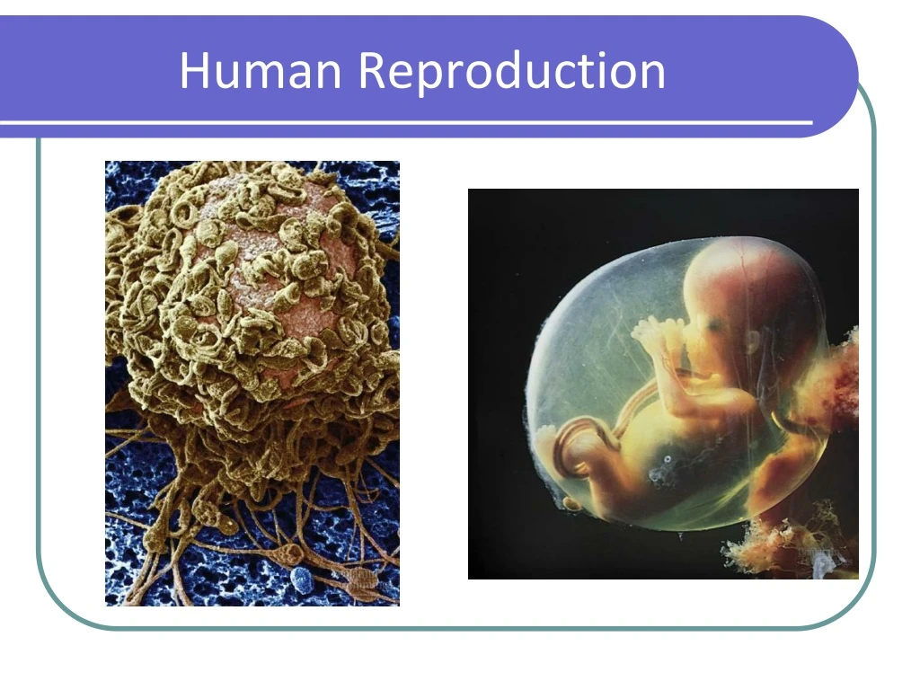
Fertilization • Fertilization is the union of a sperm and an ovum to form a diploid zygote. • Fertilization results in a zygote and triggers embryonic development. • Occurs normally inside of fallopian tube (oviduct). • Fertilization is NOT pregnancy! • (PATHWAY) Tens of millions of sperm enter the vagina cervix uterus oviducts (fertilization) sperm + ovum zygote 23(n) + 23(n) 46(2n)
Fertilization • Only one of the many millions of sperm entering the vagina will penetrate this human ovum to initiate fertilization
Fertilization • The shape of a human sperm cell is adapted to its function • Sperm cell is enzymes, DNA & mitochondria!
Multiple Fertilizations • If two (or more) ova are released in the same cycle and fertilized fraternal siblings (different DNA) • If one ova is fertilized and mitotically divides into two (or more) separate zygotes identical siblings (same DNA)
Implantation • Implantation: The fertilized zygote implants into thickened uterine lining and the embryo starts to secrete the hormone human chorionic gonadotropin (HCG) (the hormone used for pregnancy tests) • HCG keeps the corpus luteum functional and continuing to secrete progesterone. • By the third or fourth month, the placenta takes over for the corpus luteum, secreting enough estrogen and progesterone to maintain the pregnancy. • Implantation=Pregnancy!
Embryonic Development • Development: series of orderly, precise steps that transform a zygote into a multicellular embryo • Embryo ≡ early development stages of a multicellular organism • Includes: 1. cell division (mitotic) 2. cell growth 3. cell differentiation≡ altering of unspecialized mitotic embryonic cells into specialized cells, tissues,& organs
Early Embryonic Development • Cleavage is the first major phase of embryonic development • It is the rapid succession of cell divisions (Mitotic) • It creates a multicellular embryo from the zygote • NO growth • Embryonic growth cannot occur until implantation occurs ZYGOTE Blastocoel Cross sectionof blastula BLASTULA(hollow ball)
Early Embryonic Development • Stages: • Morula≡ solid ball of cells • Blastula ≡ single layer of cells surrounding a fluid-filled cavity called the blastocoel • NO growth; still dividing original single cell mass ZYGOTE Blastocoel Cross sectionof blastula BLASTULA(hollow ball)
Embryonic Development • Gastrulation is the second major phase of embryonic development • The cells at one end of the blastula move inward
Embryonic Development • Organs start to form after gastrulation • Embryonic tissue layers begin to differentiate into specific tissues and organ systems
Embryonic Membranes • Amnion ≡ fluid filled sac for protection • Chorion ≡will form the embryo’s part of the placenta • Yolk sac ≡produces first blood cells & germ cells Chorion Amnion Allantois Yolk sac
Embryonic Membranes • Allantois ≡will form the umbilical cord (ropelike structure that attaches embryo to uterus) • Umbilical cord brings nutrients in/wastes out of fetus • Ties into hepatic artery & vein in fetus - bellybutton Chorion Amnion Allantois Yolk sac