Download
1 / 21
280 likes | 1.04k Views
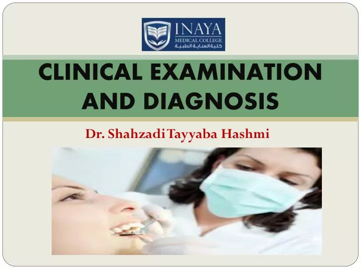
CLINICAL EXAMINATION AND DIAGNOSIS. Dr. Shahzadi Tayyaba Hashmi. CLINICAL EXAMINATION. Clinical examination: It includes both extra oral and intra oral examination. ORAL EXAMINATION AND DIAGNOSIS. Intra oral examination Hard tissue and soft tissue examination Extra oral examination

E N D
CLINICAL EXAMINATION AND DIAGNOSIS Dr. Shahzadi Tayyaba Hashmi
CLINICAL EXAMINATION • Clinical examination: • It includes both extra oral and intra oral examination
ORAL EXAMINATION AND DIAGNOSIS • Intra oral examination • Hard tissue and soft tissue examination • Extra oral examination • Head and neck examination • Face(gross abnormality) • Skin(pallor , pigmentation and cyanosis) • Eyes( anaemia and jaundice) • Nose(nasal deviations) • T M J (deviation of mandible , any mass over TMJ , tenderness on palpation, clicking sounds) • Lymph nodes of head and neck (site , size, number, consistency , tenderness ,fixity ) • Salivary gland( enlargement of major glands, dryness of mouth, quantity and character of secretion)
ORAL EXAMINATION AND DIAGNOSIS • Following sequence is followed during clinical examinations • Inspection • Palpation • Percussion • Auscultation
1) INSPECTION • Patient should be observed for : • unusual gait and habits (may suggest underlying systemic disease, drug or alcohol abuse) • Localizedswelling, • Presence of bruises, • Abrasions, scars • Signs of trauma • Degree of mouth opening, it should be at least two fingers
INSPECTION • During intraoral examination, look at the following structures systematically • Thebuccal,labialandalveolarmucosa • Thehardandsoftpalate • Thefloorofthemouthandtongue • Theretromolarregion • Theposteriorpharyngealwall andfacialpillars • Thesalivaryglandandorifices
INSPECTİON (GENERAL DENTAL STATE) • Oralhygienestatus • Amountandqualityofrestorativework • Prevalenceofcaries • Missingtooth • Presence ofsoftorhardswelling • Periodontalstatus • Presence ofanysinus tracts • Discoloredteeth • Toothwearandfacets
PALPATİON • Localriseintemperature • Tenderness • Extentoflesion • Induration • Fixationtounderlyingtissues
PERCUSSİON • Percussion gives information about the periodontal status of the tooth • Percussion of tooth indicates • inflammation in periodontal ligament which could be due to • Trauma • Sinusitis • PDLdisease
HOW CAN WE DO PERCUSSION? • Percussion can be carried out by : • gentle tapping with gloved finger • Blunt handle of mouth mirror • Each tooth should be percussed on all the surfaces of tooth until the patient is able to localize the toothwithpain.Degree of response to percussion isdirectly proportional to degree of inflammation
PERİODONTAL EVALUATİON • Periodontal examination shows change in • color • contour • form • density • level of attachment • bleeding tendency
PERİODONTAL EVALUATİON • The depth of gingival sulcus is determined by systemic probing using a periodontal probe • A sulcus depth greater than 3 mm and the sites that bleed upon probing should be recorded in the patient’s chart • The presence of pocket may indicate periodontal disease
PERİODONTAL EVALUATİON • How can we check the mobility of the tooth: • The mobility of a tooth is tested by placing a finger or blunt end of the instrument on either side of the crown and pushing it and assessing any movement with other finger
PERİODONTAL EVALUATİON • Mobility grades: • Slight (normal) • Moderate mobility within a range of 1 mm. • Extensive movement (more than 1 mm) in mesiodistal or lateral direction combined with vertical displacement in alveolus • As a general rule, mobility is graded clinically by applying firm pressure with either two metal instruments or one metal instrument and a gloved finger • Normal mobility Grade I: Slightly more than normal (<0.2mm horizontal movement) • Grade II :Moderately more than normal (1-2mm horizontal movement) • Grade III: Severe mobility (>2mm horizontal or any vertical movement)
AUSCULTATION • Intra orally of less importance • But useful in assessing movement of Temporomandibular joints