Download
1 / 22
1.63k likes | 4.64k Views
The Microscope. Curriculum Outcomes Addressed: • Explain that it is important to use proper terms when comparing plant and animal cells (109-13) • Compare the early idea that living organisms were made of air , fire, and water with the modern cell theory (110-2 )

E N D
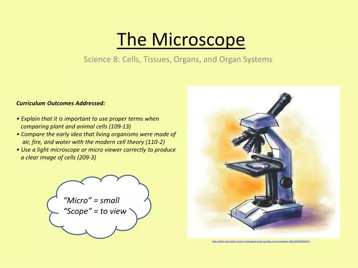
The Microscope • Curriculum Outcomes Addressed:• Explain that it is important to use proper terms when comparing plant and animal cells (109-13) • • Compare the early idea that living organisms were made of air, fire, and water with the modern cell theory (110-2) • • Use a light microscope or micro viewer correctly to produce a clear image of cells (209-3) Science 8: Cells, Tissues, Organs, and Organ Systems “Micro” = small “Scope” = to view http://office.microsoft.com/en-ca/images/results.aspx?qu=microscope&ex=2#ai:MC900382575|
Intro to the Microscope In this lesson, we will learn about… • The early idea that living organisms were made up of air, fire, and water • The two most common types of microscopes • Compound Light Microscope • Scanning Electron Microscope • The main parts of a compound light microscope • How to use a compound light microscope to produce a clear image of specimens
Key Terms • Microscope: An optical instrument used for magnifying and viewing very small objects • Magnification: The degree to which something is enlargedPower • Resolution: The amount of detail in an image • Specimen: A small quantity or sample of something that is used to represent the larger or original type of its origin (i.e., drop of blood to represent all blood)
Early Ideas of the Cell Theory • In ca. 494 BC, ancient Greek philosophers believed that everything around them was made up of four classical elements; earth, wind, fire, and water. This belief was based on the natural observations of the phases of matter around them. • Earth represented everything that was solid • Water represented everything that was liquid • Air represented everything that was gas • Fire represented everything that was plasma • The Greek Philosopher Aristotle also believed that the body was made up of four liquids(corresponding to the four elements); phlegm, blood, yellow bile and black bile, and that If a person had too much of one of these, they would fall ill. http://www.localhistories.org/science.html
Revolution of the Cell Theory • The first compound microscope was invented in the 1590s by Dutch spectacle maker Zacharias Jansen. • In 1658 Jan Swammerdan (Dutch Biologist) first observed red blood corpuscles. • In 1661 Marcello Malpighi (Italian Doctor) discovered capillaries (blood vessels). • Robert Hooke (English philosopher and architect) was the first person to describe cells in his book called Micrographia(in 1665). • Anton van Leeuwenhoek made many contributions in the world of microscopy (i.e., discovering the vacuole, and is therefore known worldwide as the “Father of Microscopy” (1723). • The modern cell theory was first proposed in the 1650s http://www.localhistories.org/science.html
Types of Microscopes There are a number of different types of microscopes. We will focus on the two most commonly used microscopes. • Compound Light Microscope (Optical Microscope) • Called “compound” because it is made up of more than one lens • Scanning Electron Microscope (SEM) • Called “scanning electron” because it electrons scan the topography of a specimen to create and magnify an image
Compound Light Microscope vs. Scanning Electron Microscope http://en.wikipedia.org/wiki/Optical_microscope http://upload.wikimedia.org/wikipedia/commons/2/2d/Transmission_electron_microscope_(Philips_TEM)_Mega_View_II_pl.jpg
Compound Light Microscope vs. Electron Microscope Image • Light Microscope Electron Microscope • 2D image - 3D image • Low resolution (blurry image) - High resolution (very clear image) http://2012books.lardbucket.org/books/general-chemistry-principles-patterns-and-applications-v1.0m/section_10_04.html
Compound Light Microscope Image of a Fruit-Fly http://www2.le.ac.uk/offices/press/press-releases/2011/june/trans-atlantic-team-announce-huntingtons-disease-breakthrough
Electron Microscope Image of a Fruit-Fly http://www.telegraph.co.uk/science/picture-galleries/7397841/Creepy-crawlies-Amazing-Scanning-Electron-Microscope-pictures-of-insects-and-spiders.html?image=3
Electron Microscope Images of a Snowflake http://en.wikipedia.org/wiki/Scanning_electron_microscopy http://en.wikipedia.org/wiki/Scanning_electron_microscopy
The Size of Cells • The majority of cells are microscopic and cannot be seen with the unaided eye (without a microscope) • 1 micrometer (µm) = one millionth of a meter Interactive Cell Size and Scale: http://learn.genetics.utah.edu/content/begin/cells/scale/
Proper Use of the Light Microscope • It is very important to follow instructions on how to carefully use a microscope. Microscopes can easily be broken, especially when it comes to the objective lenses. • Always remember to… • Carry the microscope with two hands; one hand holding the arm and one hand supporting the base • Check that the low-power objective (the smallest) is in position over the stage and no closer than 0.5cm to the stage • Turn both the coarse and the fine adjustment knobs very slowly when trying to focus on the specimen, and never allow the objective lens to touch the slide. This can break the slide and scratch or even break the objective lens http://www.deftstudios.com/bioweb/images/blab03A.gif
Determining Magnification Power • Most common light microscope eyepieces are at 10x • There are usually 3-4 objective lenses (4x, 10x, 40x, 100x) • In order to determine the magnification power (how many times you have magnified your image), you must multiply your eyepiece power (magnification) by your objective lens power (magnification). Eyepiece power X Objective lens power = Power of Magnification Example: If you are using the 4x objective lens… 10xEyepieces X 4xobjective = 40xFinal Magnification
Determining Magnification Power: Practice Q#1: If you are using the 40x objective lens on a microscope that has a 10x eyepiece, what is your magnification power?____x Eyepiece X _____x Objective lens = _______x Final Magnification Q#2: What happens to the image in view as the power of magnification increases? ______________________________
Determining Magnification Power: Practice Q#1: If you are using the 40x objective lens on a microscope that has a 10x eyepiece, what is your magnification power?10x Eyepieces X 40xobjective = 400x Final Magnification Q#2: What happens to the image in view as the power of magnification increases? __The image becomes larger__
How to use a Microscope • Step 1: Place the microscope on the table with the arm facing your body. • Step 2: Make sure that the low-power objective (smallest objective lens) is in position over the stage (facing the stage) and no closer than 0.5cm • Step 3: Rotate the diaphragm to get your optimum light • Step 4: Place your slide on the stage, adjusting it so that the specimen is directly under the lens – the specimen should be in view when you are looking through the eyepiece • Step 5: Focus on the specimen by slowly moving the coarse-adjustment knob so that the slide is being moved away from the lens. • Step 6: Rotate the revolving nosepiece so that the medium-power objective is in position. Focus with the fine-adjustment knob. • Step 7: Rotate the revolving nosepiece so that the high-power objective is in position. Focus with the fine-adjustment knob. • Step 8: When you are finished with the microscope, return to the low-power objective lens, and remove the slide from the stage.
Helpful Resources • Interactive Microscope Tutorial (Parts and Functions): http://www.microbelibrary.org/images/jones/player.html • Microscope parts and functions:http://sciencespot.net/Media/microparts.pdf • How to properly use a microscope: http://www.microscope-microscope.org/basic/how-to-use-a-microscope.htm • Microscope parts and functions quiz: http://www.purposegames.com/game/microscope-parts-functions-quiz • Interactive Microscope Online: http://www.udel.edu/biology/ketcham/microscope/scope.html