Download
1 / 33
330 likes | 338 Views
Learn about the closed system of the heart and blood vessels and how it functions in transportation, gas exchange, heat regulation, and defense. Understand the structure and function of blood vessels, capillary exchange, and the mechanisms of blood circulation. Explore the anatomy of the heart and its chambers, valves, conduction system, and regulation of heart rate. Dive into the concept of cardiac output, the regulation of the cardiovascular system, and the measurement and variations of blood pressure. Finally, discover common cardiovascular disorders like atherosclerosis and congestive heart failure.

E N D
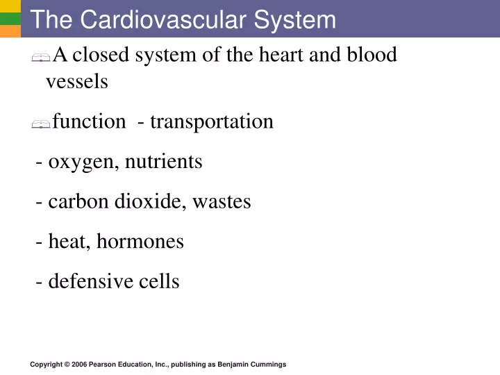
The Cardiovascular System • A closed system of the heart and blood vessels • function - transportation - oxygen, nutrients - carbon dioxide, wastes - heat, hormones - defensive cells
Blood Circulation Figure 11.3
Blood Vessels: The Vascular System • Taking blood to the tissues and back • Arteries • Arterioles • Capillaries • Venules • Veins Figure 11.8a
The Vascular System • Three layers: endothelium, middle, outer layer • Middle layer is smooth muscle Figure 11.8b
Capillary Exchange • Fluid forced out by blood pressure • Plasma proteins,blood cells retained in blood • Osmotic pressure brings fluid back into capillary • Excess tissue fluid collected into lymphatic system, returned to blood
Capillary Exchange: Mechanisms • Direct diffusion across plasma membranes • Endocytosis or exocytosis • intercellular slits = gaps • Plasma membrane not joined by tight junctions • Pores
Blood Vessels • Venous system • three layers, thin-walled • carry blood toward the heart • Mechanisms in blood return • Contraction of skeletal muscles • One-way valves • Pressure changes associated with breathing
Arterioles and Capillaries Figure 8.2
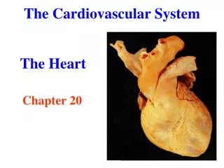
The Heart Fist-sized; placed between lungs Figure 11.1
The Heart: Heart Wall • Three layers • Epicardium • visceral pericardium • Connective tissue layer • Myocardium • Mostly cardiac muscle • Endocardium • Endothelium
The Heart: Chambers • Right and left side act as separate pumps • Four chambers • Atria • Receiving chambers • Right atrium • Left atrium • Ventricles • Discharging chambers • Right ventricle • Left ventricle Figure 11.2c
The Heart: Associated Great Vessels • Aorta • Leaves left ventricle • Pulmonary arteries • Leave right ventricle • Vena cava • Enters right atrium • Pulmonary veins (four) • Enter left atrium
The Heart: Valves • Allow blood to flow in only one direction • Four valves • Atrioventricular valves – between atria and ventricles • Semilunar valves between ventricle and artery • Valves open as blood is pumped through • AV held in place by chordae tendineae (“heart strings”) • prevent backflow
Operation of Heart Valves Fig 11.4
The Heart Figure 8.8
External Heart Anatomy Figure 11.2a
The Heart: Conduction System • Intrinsic conduction system (nodal system) • Heart muscle cells contract, without nerve impulses, in a regular, continuous way
The Heart: Conduction System • Special tissue sets the pace • Sinoatrial node • Pacemaker • Atrioventricular node • Atrioventricular bundle
Electrocardiograms (EKG/ECG) Figure 8.15b,c
Filling of Heart Chambers – the Cardiac Cycle Figure 11.6
Pulse • Pulse – pressure wave of blood • Monitored at “pressure points” where pulse is easily palpated Figure 11.16
The Heart: Cardiac Output • Cardiac output (CO) • Amount of blood pumped by each side of the heart in one minute • CO = (heart rate [HR]) x (stroke volume [SV]) • Stroke volume • Volume of blood pumped by each ventricle in one contraction
The Heart: Regulation of Heart Rate • Decreased heart rate • Parasympathetic nervous system • High blood pressure or blood volume • Decreased venous return
Cardiac Output Regulation Figure 11.7
Regulation of the Cardiovascular System • Baroreceptors: pressure receptors in aorta andcarotid arteries • BP rises, vessels stretched, signals sent to brain • Heart lowers heart rate and force of contraction • Arterioles vasodilate, increasing blood flow to tissues • Neural factors - medulla oblongata • Sympathetic nerves: constrict blood vessels • Parasympathetic nerves: dilate blood vessels • Hormones: epinephrine (adrenaline)
Measuring Arterial Blood Pressure • Diastolic – pressure when ventricles relax • Systolic – pressure at the peak of ventricular contraction Figure 11.18
Variations in Blood Pressure Normal range is variable • 140–110 mm Hg systolic • 80–75 mm Hg diastolic • Hypotension • Low systolic (below 100 mm HG) • Often associated with illness • Hypertension • High systolic (above 140 mm HG) • Can be dangerous if it is chronic
Regulation of Blood Pressure Renal factors (kidneys) - alter blood volume Renin – stimulates hormonal control
Blood Pressure: Effects of Factors • Temperature • Heat - vasodilation effect • Cold - vasoconstricting effect • Chemicals • Various substances can cause increases or decreases • Diet
Atherosclerosis • Buildup of plaques on inner wall of arteries • Source - injury from chemicals, physical blow or pressure. injured cells attract lipid deposits, muscle cells which block lumen of vessel Arteriosclerosis - hardening, loss of elasticity in wall as cells die, fibers degenerate, rigid scar tissue replaces elastic cells.
Cardiovascular Disorders • Angina pectoris: a warning • Myocardial infarction (heart attack): permanent cardiac damage • Congestive heart failure: decrease in pumping efficiency • Embolism: blockage of blood vessels • Stroke: impaired blood flow to the brain
Reducing the Risk of Cardiovascular Disease • Smoking: don’t • Blood lipids: monitor cholesterol levels • Exercise: regular and moderate • Blood pressure: treat hypertension • Weight: being overweight increases risk of heart attack and stroke • Control of Diabetes Mellitus: early diagnosis and treatment delays onset of related problems • Stress: avoid chronic stress