Download
1 / 60
600 likes | 607 Views
Explore the respiratory system anatomy, terminology, and structural plan including upper respiratory tract organs like the nose and pharynx. Learn about respiratory processes, cellular respiration, and common respiratory system disorders.

E N D
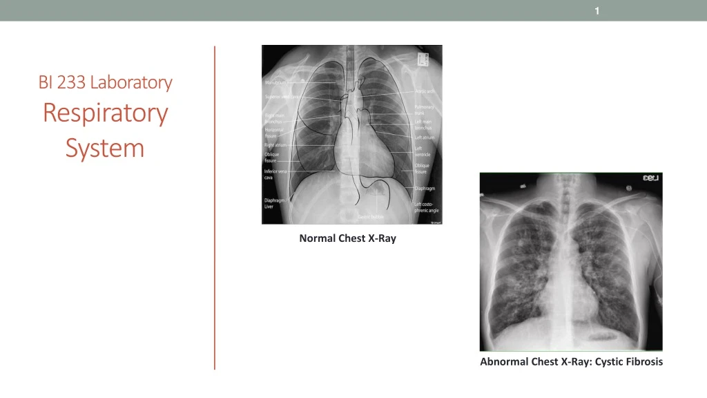
BI 233 Laboratory RespiratorySystem Normal Chest X-Ray Abnormal Chest X-Ray: Cystic Fibrosis
Organs of the Respiratory System • Nose • Pharynx • Larynx • Trachea • Bronchi • Lungs • Bronchi • Bronchioles • Alveoli Kahn Academy video on respiratory anatomy
Function of the Respiratory System • Cells continually use O₂ and release CO₂. • Gas exchanges between blood and external environment. • Occurs in alveoli of lungs. • Cardiovascular system transports gases in blood. • Passageways to lungs purify, humidify, and warm incoming air. • Failure of either system… • Rapid cell death from O₂ starvation.
Respiratory Terminology • Pulmonary ventilation • Process by which O₂ enters and CO₂ exits alveoli (breathing). • Respiration • Process by which O₂ and CO₂ diffuse in and out of the blood (gas exchange) • Occurs in 2 areas of the body. • External respiration – gas exchange across respiratory membrane of lungs (alveoli) into the blood. • Internal respiration – gas exchange between the blood and the interstitial fluid, tissues and cells • Cellular respiration • Series of metabolic processes where living cells produce energy through oxidation of organic substances.
Structural Plan of the Respiratory System • Airflow in lungs. • Bronchi bronchioles alveoli • Conducting division • Passages serve only for airflow, nostrils to bronchioles.. • Respiratory division • Alveoli and distal gas-exchange regions. • Two structural divisions • Upper respiratory tract • Organs in head and neck, nose through larynx. • Lower respiratory tract • Organs of the thorax, trachea through lungs. • Accessory structures • Include: oral cavity, rib cage, and diaphragm.
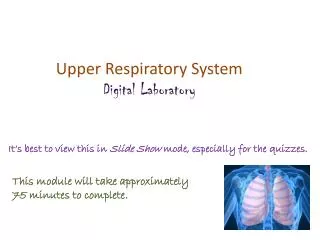
Upper Respiratory Tract:Organs in head and neck, nose through larynx.
Nose • Functions • Provides a passage way for air travel to and from lungs. • Warms, cleanses, humidifies inhaled air. • Makes possible the sense of smell. • Resonating chamber that amplifies the voice. • Bony and cartilaginous supports • Superior half • Nasal bones medially maxillae laterally. • Inferior half • Lateral and alar cartilages. • Ala nasi • Flared portion shaped by dense CT, forms lateral wall of each nostril.
Nose:Extends anteriorly from nares to posterior nasal apertures. • Nares (nostrils) • External openings of the nasal cavity. • Ethmoid and sphenoid bones compose the roof. • Palate forms the floor. • Vestibule • Dilated chamber inside ala nasi. • Stratified squamous epithelium, vibrissae (guard hairs). • Nasal septum • Divides cavity into right and left chambers called nasal fossae. • Formed by perpendicular plate of ethmoid bone and vomer.
Nasal Conchae • Superior, middle and inferior nasal conchae. • 3 folds of tissue on lateral wall of nasal fossa. • Mucous membranes supported by thin scroll-like. turbinate bones. • Meatuses • Narrow air passage beneath each conchae. • Narrowness and turbulence ensures air contacts mucous membranes.
Mucosa: Pseudostratified Columnar Ciliated Epithelium • Olfactory mucosa lines roof of nasal fossa. • Respiratory mucosa lines rest of nasal cavity. • Pseudostratified columnar ciliated epithelium (PCCE). • Defensive role of mucosa. • Mucus (from goblet cells) traps inhaled particles. • Bacteria destroyed by lysozyme. • Cilia • Drive debris-laden mucus into pharynx to be swallowed or expectorated.
Paranasal Sinuses:Cavities within Bones Surrounding Nasal Cavity • Air conducting spaces that open and drain into nasal cavity. • Lined with respiratory epithelium. • Located in the following bones: • Frontal bone • Sphenoid bone • Ethmoid bone • Maxillary bone • Function • Lighten the skull. • Act as resonance chambers for speech. • Produce mucus that drains into the nasal cavity.
Pharynx:Pathway for Respiratory and Digestive Tract • Three distinct regions: • Nasopharynx • PCCE - Pseudostratified columnar ciliated epithelium tissue. • Posterior to nasal cavity, dorsal to soft palate. • Receives auditory tubes and contains pharyngeal tonsil. • Oropharynx • SSE - Stratified squamous epithelium. • Space between soft palate and root of tongue. • Common passageway for food and air. • Contains palatine and lingual tonsils. • Laryngopharynx (hypopharynx) • SSE - Stratified squamous epithelium. • Hyoid bone to cricoid cartilage (inferior end of larynx).
Auditory Tube • AKA: Eustachian tube • Links middle ear to nasopharynx. • Function is to protect, aerate and drain middle ear (and mastoid). • Occlusion leads to development of middle ear inflammation (otitis media).
Tonsils • Tonsils are lymphoid tissue. • First line of defense of nasal and oral cavities. • Pharyngeal (adenoid) • Palatine • Lingual
Uvula:Helps close off nasopharynx. • Small, fleshy, conical body projecting downward from middle of soft palate. • During swallowing • Soft palate elevates and uvula helps closes off nasopharynx. • Prevents food and fluids from entering nasal cavity.
Larynx:Forms Portion of Airway to Lung and Produces Phonation • Positioned between root of tongue and upper end of trachea. • Structures • Epiglottis • Flap of tissue guarding glottis, directs food and drink to esophagus. • Elastic cartilage. • Glottis • Superior opening. • Consists of vocal cords AND rima glottis (glottidis). • Swallowing Video
Cartilage and Ligaments of the Larynx • Thyroid cartilage (“Adam’s apple”/hyaline cartilage) • Cricoid cartilage (hyaline cartilage) • Cricothyroid ligament • Site of emergent cricothyrotomy. • Thyrohyoid membrane • Tracheal cartilage (hyaline cartilage) • Hyoid bone Thyrohyoid membrane
Structure of the Larynx Continued • Interior wall • Two folds on each side, from thyroid to arytenoid cartilages. • Vestibular folds (false vocal cords/upper) • Superior pair, close glottis during swallowing. • Vocal Folds (true vocal cords/lower) • Produce sound. • Space between vocal folds is rima glottis. • Intrinsic muscles • Insert and originate on larynx. • Rotate corniculate and arytenoid cartilages. • Adducts (tightens) - high pitch sound. • Abducts (loosens) - low pitch sound. • Extrinsic muscles • Connect larynx to hyoid bone, elevate larynx during swallowing.
Speech and Whispering • Speech • Modified sound made by the larynx. • Requires pharynx, mouth, nasal cavity and sinuses to resonate sound. • Tongue and lips form words. • Pitch is controlled by tension on vocal folds. • Pulled tight produces higher pitch. • Male vocal folds are thicker and longer. • Vibrate more slowly producing a lower pitch. • Whispering is forcing air through almost closed rima glottidis. • Oral cavity alone forms speech.
Larynx Videos https://www.youtube.com/watch?v=mJedwz_r2Pc https://www.youtube.com/watch?v=-XGds2GAvGQ
Lower Respiratory Tract:Organs of thorax, trachea through lungs. Carina Left main (primary) bronchus
Trachea • Rigid tube 4.5 in. Long and 2.5 in. In diameter. • Anterior to esophagus. • Supported by 16 to 20 c-shaped cartilaginous. rings • Opening of rings posterior towards esophagus. • Trachealis musclespans opening in rings. • Adjusts airflow by expanding or contracting. • Larynx and trachea lined with PCCE. • Functions as mucociliary escalator. • Esophagus lies posterior to and parallels the trachea.
Reestablishing airflow past an airway obstruction. • Crushing injury to larynx or chest. • Swelling that closes airway. • Vomit or foreign object. • Tracheostomy • Incision in trachea below cricoid cartilage if larynx is obstructed. • Intubation • Passing a tube from mouth or nose through larynx and trachea. Clinical Correlation: Tracheostomy and Intubation
Carina and Primary (main) Bronchi • Carina • Ridge separating openings of right and left main bronchi at their junction with trachea. • Right and left primary (main) bronchi • Passage of airway in respiratory tract that conducts air into lungs. • Bronchi branch into smaller secondary and tertiary bronchi which again branch into smaller bronchioles. • No gas exchange takes place in bronchi. right main (primary) bronchus
Apex, Base and Hilum of the Lung • Occupy most of the thoracic cavity. • Heart occupies central portion called mediastinum. • Apex • Superior portion. • Near the clavicle. • Base • Inferior portion. • Rests on diaphragm. • Hilum (hilus) • Slit on lungs medial surface where primary bronchi and pulmonary blood vessels enter.
Lobes of the Lungs • Each lung is divided into lobes separated by fissures. • Right lung • Three lobes: superior, middle, and inferior. • Superior and middle separated by horizontal fissure. • Middle and inferior separated by oblique fissure. • Left lung • Due to heart only included two lobes: superior and inferior. • Superior lobe includes cardiac notch (impression) accommodating heart. • Superior and inferior separated by oblique fissure.
Pleura, Pleural Cavity, and Pleural Fluid • Pleurae and its layers. • Visceral pleura • Covers lungs. • Parietal pleura • Lines ribcage and upper surface of diaphragm. • Pleural cavity and serous fluid. • Potential space between pleural layers with capacity to produce serous fluid. • Functions of serous fluid… • Reduction of friction. • Creation of pressure gradient. • Lower pressure assists in inflation of lungs. • Compartmentalization • Prevents spread of infection
Bronchi of the Lung • Primary bronchi • Arise from trachea, after 2-3 cm enter hilum of lungs. • Divide into two primary bronchi ( right and left). • Right bronchus slightly wider and more vertical. • Most common site for aspiration of foreign objects. • Secondary (lobar) bronchi • Primary bronchi enter lungs and divide into secondary bronchi. • Overlapping plates. • Branches into one secondary bronchus for each lobe. • Tertiary (segmental) bronchi • Overlapping plates. • 10 right, 8 left. • Bronchopulmonary segment: portion of lung supplied by each.
Bronchioles and Alveoli of the Lung • Bronchioles • Lack cartilage. • Have layer of smooth muscle. • Pulmonary lobule: portion ventilated by one bronchiole. • Divides into 50 - 80 terminal bronchioles. • Terminal bronchioles • Have cilia , give off two or more respiratory bronchioles. • Respiratory bronchioles • Divide into 2-10 alveolar ducts. • Alveolar ducts • End in alveolar sacs. • Alveoli • Bud from respiratory bronchioles. • Alveolar ducts and alveolar sacs.
Alveoli Histology • Epithelium transitions from PCCE to non-ciliated simple cuboidal, and finally, moving deeper into lungs, to simple squamous epithelial tissue allowing for effective gas exchange.
Cell Types of Alveoli • Type I alveolar cells • Simple squamous cells where gas exchange occurs • Type II alveolar cells • Septal cells • Free surface has microvilli. • Secrete alveolar fluid containing surfactant. • Alveolar dust cells • Wandering macrophages remove debris.
Alveolar-Capillary Membrane • Respiratory membrane • 1/2 micron thick. • Exchange of gas from alveoli to blood. • Four layers of membrane to cross. • Alveolar epithelial wall of type I cells. • Alveolar epithelial basement membrane. • Capillary endothelial basement membrane. • Endothelial cells of capillary-> blood • Vast surface area equivalent to size of handball court.