Download
1 / 30
310 likes | 476 Views
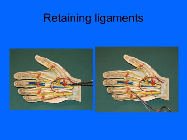
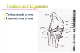
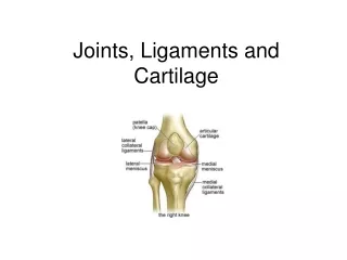
Ligaments. PTP 521 Musculoskeletal Disorders and Dysfunctions. I. Ligaments. Latin: Ligare : to tie or bind Named: Bones in which they insert Shape Relationship to joints Relationship to each other. Ligaments. 1. Connect two bones together 2 . Stabilize joints

E N D
Ligaments PTP 521 Musculoskeletal Disorders and Dysfunctions
I. Ligaments • Latin: Ligare: to tie or bind Named: • Bones in which they insert • Shape • Relationship to joints • Relationship to each other
Ligaments 1. Connect two bones together 2. Stabilize joints 3. Connective tissue structure 4. Shared function of restraint 5. Proprioceptor for the joint
Histological Composition of Ligaments • 2/3 water • Contributes to cellular function • Viscoelastic behavior • 1/3 solid • Collagen (up to 27 types have been identified in ligaments, generally considered to be one of 6 types • Proteoglycans – control collagen fibrillogenesis by controlling fibril diameter and the rate of fibril formation • Elastin: <2% of most ligaments
Influence of Hormones on Ligaments • Some ligaments have receptors for hormones (estrogen, progesterone, and androgens) • Ligament function could be regulated by hormones • Might be gender-specific of regulation
Evidence to Support Hormonal Influence: • During pregnancy, hormone relaxin is released to cause laxity in ligaments • Women more likely to have benign joint hypermobility syndrome • Ligamentous laxity during menstrual cycle – not clearly defined
Effects of Immobilization • Load Deprivation: rapid deterioration in biochemical and mechanical properties • Ligaments are strain-rate sensitive • Stronger and stiffer at higher loading rates
Decrease in ligament mass causes net loss in ligament strength and stiffness • Shift in ligament cell metabolism from a building or steady state to a destructive state • Few weeks of immobility: ligament matrix quantity decreases • Bone will resorb causing weakness at insertional sites
Immobilization greater than 6-9 weeks can result in as much as a 50% reduction in strength
Effects of Exercise • Response to exercise: increase strength and stiffness (minimally) • Potential to increase ligament strength and stiffness no more than 10-20% • Effects of exercise may be ligament specific
Ligament Injuries • One of the most common injuries to joint • MOI: Falls, twisting, getting hit • Knee in particular is susceptible • 25-40%
Injury to Ligaments • Sprain a. Definition: disruptions of fibers of a ligament b. Key point • Caused by a force that stretches fibers beyond elastic limits
Mechanisms of Injury • Joint Dependent • Extrinsic Load applied to joint • Ligaments parts that are the best to restrain in that position are loaded the most • Deform past elastic limit • Fail
Force required and amount of instability occurring are dependent upon • 1) size of ligament • 2) age of person • 3) position of joint when force occurs
Failure occurs at insertion, body of ligament or bone interface (avulsion) • Children • Adults • Evaluate immediately after an injury
Unstable only in the position of injury. • True of partial ligament tears only • MRI, stress radiographs and arthrograms are all imaging studies to determine the existence of a ligament sprain
Avulsion Fractures • Force: tensile loading of tissue • Causes insertion site to pull away from bone taking a part of bone with it
Enthesitis • Definition: inflammation of insertion of ligament or tendon • Extra-articular • Younger child may be called apophysitis
Ligament Sprains • Classification of Sprains: • Number of fibers disrupted • Instability of the joint involved
Grade I: mild or first degree sprain Signs: ROM limited in one direction only • Increase in pain during PROM and AROM – when ligament is stressed • Limited bruising Symptoms: localized tenderness over the injury area, pain
Grade II: moderate or second degree sprain Signs: clinical evidence of instability • Stress testing gives slight laxity but not complete functional instability • Ecchymosis • Decrease in ROM, painful, swelling restricts movement • Edema Symptoms: more diffusely tender • Increase in pain • Instability with weight bearing and with movement that would stress the ligament • Generally parts of several ligaments are injured with a second degree
Grade III: severe or third degree sprain Signs: significant laxity is seen, abnormal increase in ROM • Severe swelling • Ecchymosis or hemarthrosis • Structural, functional instability Symptoms: may be less painful than a grade II sprain May also have second degree sprains in other ligaments
Joint Instability Stress testing: compare the injured and the non-injured side 0 instability: No difference between injured and non-injured side 1+ instability: difference is less than 0.5 cm 2+ instability: difference is between 0.5cm and 1cm 3+ instability: difference is greater than 1cm
Ligamentous Healing Phase 1: Inflammation, occurs within 72 hours. • Inflammatory response • Immediate pain and bleeding • Extra-articular ligaments: bleeding outside joint cavity • Intra-articular ligaments: bleeding within joint cavity
Phase 2: Repair/Regeneration: 48-72 hours after injury and lasts up to 6 weeks • Production of scar matrix, viscous material becomes less viscous as days go on
Healing Failure: a. failure to reconnect appropriate locations on bones b. failure to produce enough scar tissue c. failure to produce the correct type of tissue
Phase 3: Remodeling: 6 weeks to 12 or more months to complete • Over time, becomes more ligamentous, some differences exist in both composition and architecture of the ligament.
Surgical Repair Guidelines 1. Patient is high risk for instability in the future 2. Age of patient 3. Type of ligament sprain that occurs 4. Associated lesions, degree of laxity
Types of Surgeries • Repair: reattachment of torn ends of a ligament • Reconstruction: replacement of original ligament with a graft • Autograph • Allograph