Download
1 / 12
200 likes | 569 Views
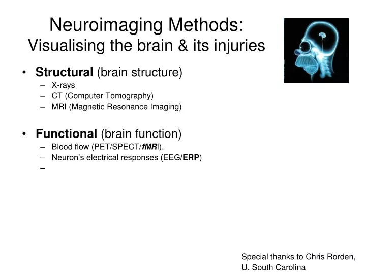
Neuroimaging Methods: Visualising the brain & its injuries. Structural (brain structure) X-rays CT (Computer Tomography) MRI (Magnetic Resonance Imaging) Functional (brain function) Blood flow (PET/SPECT/ fMR I). Neuron’s electrical responses (EEG/ ERP ).

E N D
Neuroimaging Methods:Visualising the brain & its injuries • Structural (brain structure) • X-rays • CT (Computer Tomography) • MRI (Magnetic Resonance Imaging) • Functional (brain function) • Blood flow (PET/SPECT/fMRI). • Neuron’s electrical responses (EEG/ERP) Special thanks to Chris Rorden, U. South Carolina
Structural: X-rays • X-ray tube projects through head • Detector plate measures transmission of X-rays • Bone relatively opaque to X-rays • Soft tissue relatively transparent • Use: • broken bones • Angiography • Not good for much else
Structural: CT scans Uses: • Stroke • Brain tumors (larger than 2-4 mm) • Enhanced with contrast material • Subdural Hematoma • Evaluation of traumatic Head Injury
MRI • Magnetic resonance imaging • Does not expose individual to X-rays
MRI scans Healthy enlarged ventricles MCA infarct & wide sulci
Types of MRI scan • T1 (anatomical): • fast to acquire, • good detail (e.g. white and gray matter). • T2 (pathological): • slower to acquire, thus worse resolution. • Excellent for finding lesions.
Functional imaging: Measures brain activity • PET(Positron Emission Tomography • SPECT(Single Photon Emission Computerized Tomography) • Radioactive oxygen isotope injected into blood • Brain regions that use oxygen emit more positrons
functional: fMRI • fMRI: Functional Magnetic Resonance Imaging • Increase activity in certain brain area • Blood vessels dilate • The % of Oxygen in the blood in that area is changed • The MR machine registers that
Functional: Electroencephalogram (EEG) • Measures electrical activity • When neurons fire, they create electical dipoles. • Neurons aligned perpendicular to cortical surface. - +
+ neutral Signal V _ ‘rape’ 0 100 200 300 Time (ms) Event related potentials (ERPs) • ERPs are a type of EEG • Continuously collect EEGs • Present many trials of stimuli (words: neutral vs. offensive) • Compute average brain response to stimuli • Good temporal resolution (when activity starts happening). • Poor Spatial resolution http://brainserver.psych.indiana.edu/
In sum, • Structural (brain structure) • X-rays • CT (Computer Tomography) • MRI (Magnetic Resonance Imaging) • Functional (brain function) • Blood flow (PET/SPECT/fMRI). • Neuron’s electrical responses (EEG/EEG) • Neuron’s magnetic responses (MEG)