Download
1 / 91
910 likes | 946 Views
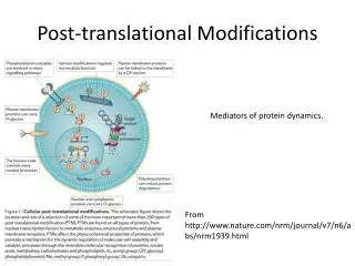
Discover the intricacies of post-translational modifications in proteins and how they impact mass spectrometry analysis. Learn about enzymatic processing, glycosylation, phosphorylation, acylation, cross-linkage, oxidation, methylation, and ubiquitination. Explore the challenges in sample preparation, mass spectrometry techniques, and assigning modifications accurately. Uncover the significance of disulfide bridges and glycosylation diversity in protein analysis. This comprehensive guide provides insights into identifying and analyzing post-translational modifications for advanced proteomic studies.

E N D
Post-translational modificationsPC235Katalin F. Medzihradszkyfolkl@cgl.ucsf.edu Post-translational Modification of Proteins Expanding Nature’s Inventory (2006) by C.T. Walsh ISBN 0-9747077-3-2
Just for introduction Genome ↓ Transcription – mRNA ↓ Translation – proteins + co-translational modifications ↓ Post-translational modifications
Post-translational modifications I • Enzymatic processing • N-, O-, C-linked glycosylation – Asn, Ser/Thr/Hyl/Hyp, Trp • Phosphorylation – Tyr, Ser, Thr, His, Asp • Acylation acetylation of the N-terminus fatty acid anchors on Cys • Cross-linkage – Lys, Trp, Tyr, Met • Oxydation – Cys, Met, Trp, Tyr, His • Methylation – N-terminus, Arg, Lys • Ubiquitination - Lys
Post-translational modifications II • Cannot be predicted – though consensus sequences have been reported for some of them • Organism-dependent • Can be tissue- or location-specific • Stable or dynamic – high and low level • Alters biological activity, and physical properties • May alter the immune response ↓
Sample preparation • Will the modification survive? • Can I get it down to a mass spectrometry friendly size? • Isolation/enrichment? • Losses – too hydrophilic/hydrophobic
Mass spectrometry • Will the modification survive the ionization? the MS/MS activation? • How unambiguous is the assignment? modification assignment? Ac vs. Me3; phosphate vs. sulfate etc. site assignment?
Finding the N-termini • 80-90% of the eukaryotic proteins is acetylated If 2nd aa: G, A, S, C, T, P, V • Met-1 is clipped off; Ac- is added to G, A, S, T If 2nd aa: E, D, Q, M, I, L, W, F – persisting Met gets acetylated
Finding the N-termini • McDonald L, et al., Positional proteomics: selective recovery and analysis of N-terminal proteolytic peptides.Nat Methods. 2005 Dec;2(12):955-7. Epub 2005 Nov 18.
Modified N-terminus Modified side-chain I don’t think any search engine will tell the difference !
Methylation (mono, di, tri - +14, +28, +42 Da) • N-terminus, Lys, Arg, His • Trimethyl – acetyl = 36 mmu Accurate mass measurement helps; Fragmentation is different too • Glu (Asp) may form Me-ester – upon CBB staining (MeOH + acid) + 14 Da
Disulfide-bridges • in membrane and secreted proteins important 3D structure feature • prone to shuffling @ basic pH
Assigning disulfide-bridges I Reduction/alkylation Reduction/alkylation m/z I Digestion @ low pH Digestion @ low pH m/z I Reduction/alkylation MS/MS I Reduction/alkylation m/z I m/z m/z
Synthetic Ac-TIMP-1(Ser175)126-184 ECTVFPCLSIPCKLQSGTHCLWTDQLLQGSEKGFQSRHLACLPREPGLCS WQSLRSQIA Where are the disulfide bridges? * digestion with trypsin @ pH 6; * with pepsin in acid Bodi, N. et al., J. Pept. Sci.9, 430-441 (2003).
[33-37] 41-49 [38-44]-[45-55] intensity [38-44] Ac[1-13]-[45-55] 2-7, 12-49 20-41 [45-55] [14-32]-[38-44] free SH 41-49 [38-55] Free SH 600 800 1000 1200 2000 2200 2400 2600 2800 3000 m/z m/z S* [45-55] 2 S* [38-55] 3 S* Ac[1-13] intensity 1 S* [14-37] 1 S* [14-32] Ac[1-13] 1 S-S, 1 S* Ac[1-13] 1free SH, 2 S* 2 S* S* [38-59] [38-44] 1 S* [33-37] [45-59] 1600 1800 2000 2200 2400 2600 2800 3000 600 800 1000 1200 1400 1600 m/z m/z MALDI-TOF analysis of the tryptic digest In-source reduction PSD
Results from the MALDI-TOF MS Tryptic digest After reduction/alkylation
Disulfide bridges in TIMP-1 C-terminal domain: [38-44]-S-S-[45-55], PSD of MH+ at m/z 2082.1 MALDI-PSD/CID yields characteristic triplets
Disulfide Bridges The ProteinProspector mass modification search can be used in conjunction with MS-Bridge to find peptides with disulfide bridges. For this example, mass shifts between 0-2000 Da were considered. (1023.3276+4). KLSWADLIVFAGNCALESMGFK+4 VSFADLVVLGGCAAIEK
Glycosylationhttp://glycores.ncifcrf.gov/Reference: Essentials of Glycobiologyby Varki et al.
N-linked AsnXxxSer/Thr/Cys
N-linked glycosylation • consensus sequence • GlcNAc2Man3 – core oligomannose structure – just Man units complex sugars– GlcNAc-Gal–SA antennae hybrid structures core fucosylation sulfate, phosphate modifications • PNGase F removes all N-linked structures; Asn Asp
N-linked glycosylation • Incredible heterogeneity: a site may be only partially occupied and may display numerous different carbohydrates • species-, tissue-, cell-type-specificmodification, physiological changes, diseasesmay alter the sugars
certain structures are immunogenic Gal a1-3 capping, Fuc a1-3 on inner GlcNAc; blood group determinants
N-linked glycosylation • Identification from diagnostic fragments: * HexNAc m/z 204 * HexHexNAc m/z 366 Precursor scan, or „ping-pong” acquisition • Identification from oligosaccharide heterogeneity • enrichment by HILIC or lectin-chromatography
human lecithin:cholesterol acyltranferase and apolipoprotein D, tryptic digest, LC/MS analysis
Recombinant Factor VIII, 50 kDa subunit MHTVNGYVN*R
AG(Man8GlcNAc2)NVSNIIPASATLNADVR peptide+GlcNAc
About the structures of N-linked glycopeptides • from the measured mass, and the CID spectrum the modified peptide can be identified + the size and class of the sugar • the identity of the sugar units and their linkage positions CANNOT be determined • NMR, exo- and endoglycosidases are needed to complete the job
[MHNa2]3+ of QV(Man10GlcNAc2)NIT and [MH2Na] 3+ of QV(Man9GlcNAc2)NITGK
One component from the previous slide K.F. Medzihradszky Meth. Enzymol.405, 116-138 (2005).
O-linked sugars • No consensus sequence • No common core structure • No universal enzyme b-elimination works (NaOH) sugars have to be reduced upon release Detection is problematic – because of heterogeneity; variable site occupancy Site assignment is even harder
Other O-linked core structures • Fuc Harris, R.J. & Spelmann, M.W. (1993) Glycobiology,3, 219-224. • Glc Nishimura, H et al., (1989) J. Biol. Chem.264, 20320-20325. • Man – in yeast ____________________________________ • GlcNAc – single unit; INSIDE the cell
J. Am. Soc. Mass Spectrom. 7, 1996, 319-328. CID fragmentation of O-linked glycopeptides
GlcNAc GlcNAc protein GlcNAc O-GlcNAcylation O-GlcNAc PSVPVSerGSAPGR WGA O-linked GlcNAc • Regulatory modification of nuclear and cytoplasmic proteins • Poorly understood due to lack of effective methods for enrichment and detection. • The Enrichment Problem • WGA lectin has affinity for GlcNAc, but affinity to a single GlcNAc moiety is low: millimolar1. Complex glycosylation Good recovery of glycoprotein Load Wash Elute WGA No recovery Load Wash Elute Ohlson S et al. Bioseparation (1998) 7 p.101
UV absorbance 214 O-GlcNAc modified enriched Non-enriched Time (minutes isocratic HPLC) O-GlcNAc Enrichment • Selective Enrichment of O-GlcNAc modified peptides using lectin weak affinity chromatography1. Peptide Mixture Isocratic Chromatography fraction collection WGA column Vosseller, K. et al. Mol Cell Proteomics (2006) 5 5: p.923-934
CID Analysis of O-GlcNAc-Modified Peptides • O-glycosidic link is significantly more labile under CID conditions than peptide backbone. • Modification site identification using CID often not possible. Chalkley, R. J. and Burlingame, A. L. J. Am. Soc. Mass Spectrom. (2001) 12 p.1106-1113
A bit about MS/MS alternatives • ECD (electron-capture dissociation) – multiply charged ions meet electron beamin FT-ICR – larger the charge state larger the capture’s efficiency • ETD (electron-transfer dissociation) – multiply charged ions meet stable anion (fluoranthene) in ion traps • radical ion is formed, different mechanism mostly backbone cleavages
Bakken, V.; Helgaker, T.; Uggerud, E., Eur. J. Mass Spectrom. 2004, 10, 625-638 Mechanism of ECD II
ECD MS Spectrum of GlcNAc-modified Peptide from Spectrin Vosseller, K et al. Mol Cell Proteomics (2006) 5 5: p.923-924
PhosphorylationBiological significance • One of the most important regulatory events: • turns proteins on and off • induces or prevents other post-translational modifications in the same protein • signaling pathways : phosphorylation cascades
Difficulties • Dynamic process : kinase vs. phosphatase • – both must be blocked during isolation • Phosphorylation often @ low level (<5%) • Lower ionization efficiency – signal of phosphopeptides suppressed Enrichment is a must at protein level at peptide level
A whole cell lysate with 20,000 sites of phosphorylation at 1% stoichiometry
A whole cell lysate with 20,000 sites of phosphorylation at 1% stoichiometry
phosphopeptide relative distribution after a 10,000 fold enrichment