Download
1 / 34
340 likes | 945 Views
Imagenes en Torax. Rx de ToraxTACEcograf

E N D
1. Monitoreo Respiratorio Radiol�gico Dr. Jorge CABRERA VALENTIN
Medicina Intensiva
UCI Hospital Alberto Sabogal
jocava26107@yahoo.com
2. MRI not widely used but can show malignancy especially
We will look at actual films in the tutorials/practicalsMRI not widely used but can show malignancy especially
We will look at actual films in the tutorials/practicals
3. Genralidades Rx de Torax Investigaci�n radiologica m�s com�n - 40% de todas las investigaciones -.
Componente estandar de un examen pulmonar
Revision sistematica es vital en la interpretacion de la Rx de Torax
Up to 40% off all radiological investigations are chest x-rays
60% are carried out in the ICU, Sensitivity; 50% of critically ill patients in Icu will have abnormal chest x-ray
A systematic review is vital when interpretating x-rays however what order is not critical and you will come across varying ordersUp to 40% off all radiological investigations are chest x-rays
60% are carried out in the ICU, Sensitivity; 50% of critically ill patients in Icu will have abnormal chest x-ray
A systematic review is vital when interpretating x-rays however what order is not critical and you will come across varying orders
4. Limitaciones de una Rx de torax Imagen de 2 dimensiones de una estructura de 3 dimensiones
Hallazgo en Rx pueden preceder a otras caracteristicas clinicas.
Una Rx normal no descarta otra patolog�a
Depende de una buena calidad de imagen A chest x-ray forms a piece in the pulmonary examination, should refer to previous x-rays if available and if possible put in context of the other pulmonary findingsA chest x-ray forms a piece in the pulmonary examination, should refer to previous x-rays if available and if possible put in context of the other pulmonary findings
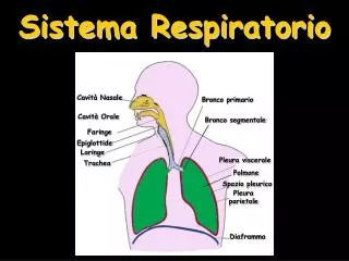
5. Vistas/tipos de Rx de Torax Posteroanterior - PA
Anteroposterior - AP
Lateral
Decubito
6. Vista PA
Servicio de Radiolog�a, estandar
Rayos X van de posterior a anterior
Posicion parado
PA
Standard investigation carried out in the x-ray dept
Cassette anterior to chest, x-rays shot post-ant from 2 metres away, shoulders abducted to remove scapula
Carried out in standing therefore better inspiration
PA
Standard investigation carried out in the x-ray dept
Cassette anterior to chest, x-rays shot post-ant from 2 metres away, shoulders abducted to remove scapula
Carried out in standing therefore better inspiration
10. Anatomia Pulmonar Transverse fissure � 6th rib laterally
Does not estend beyond pulm artery medially
Visible in 50%Transverse fissure � 6th rib laterally
Does not estend beyond pulm artery medially
Visible in 50%
12. Vista AP
Chasis colocado detras del paciente
Rayos X van de anterior a posterior
Sentado en una silla, semisentado o supino en cama,
AP marcado en la Rx
Corazon agrandado, pobre inspiraci�n AP
Cassette placed behind the patient, portable machine
Patient could be sitting in a chair, semi erect in bed, supine in bed. NOTE the patient position will affect the CXR
Marked AP on film
Heart enlarged often poorer expansion
AP
Cassette placed behind the patient, portable machine
Patient could be sitting in a chair, semi erect in bed, supine in bed. NOTE the patient position will affect the CXR
Marked AP on film
Heart enlarged often poorer expansion
13. Paciente en UCI, Limitaciones:
La calidad de la Rx puede variar ampliamente: de buena a no interpretable
Incapacidad de cooperar
La naturaleza de los ambientes de UCI
Dificultad en controlar la radiaci�n en obesos
Amplias diferencias en la exposici�n
Se sugiere estandarizar la tecnica: posicion supina, placa vertical, distancia 120 cm, inspiracion pico, usando de 80 a 100 KVp y corto tiempo de exposicion para minimizar el artefacto respiratorio
Cables de monitoreo y otros objetos externos deber�an ser removidos
14. Guia para ver una Rx Torax Rx Correcta
Prepracion
Cuarto oscuro
Placas previas si estan disponibles
Identificacion: Nombre, Fecha, Hora
Tipo : PA, AP, supino, parado
Calidad: Rotaci�n, lordosis, penetracion, inspiracion
Distancia, potencia, tiempo de exposcion.
Aparatos invasivos: Tubos, CVC, cables, How to view
Check patient and x-ray details
Left or right, markers placed on by radiographer, stomach on left. Heart not always on leftHow to view
Check patient and x-ray details
Left or right, markers placed on by radiographer, stomach on left. Heart not always on left
15. Guia para ver una Rx Torax Tejidos blandos y oseos
Cuello, supraclavicular, axila, pared torax, mama, burbuja gastrica
Humero, union escapular, escapula, clavicula, vertebra, costillas y esternon
Mediastino
Superior, medio
Tama�o, forma, densidad
Corazon
Tama�o, forma, silueta
How to view
Check patient and x-ray details
Left or right, markers placed on by radiographer, stomach on left. Heart not always on leftHow to view
Check patient and x-ray details
Left or right, markers placed on by radiographer, stomach on left. Heart not always on left
16. Guia para ver una Rx Torax Diafragma
Forma, angulo costofrenico
Pleura/Cisuras
Pulmones
Traquea y bronquios
Hilio
Vasculatura
Parenquima
Apex
Detras del coraz�n
How to view
Check patient and x-ray details
Left or right, markers placed on by radiographer, stomach on left. Heart not always on leftHow to view
Check patient and x-ray details
Left or right, markers placed on by radiographer, stomach on left. Heart not always on left
17. Basico Im�genes Radiolucidas
Im�genes Radiopacas
18. Aparatos invasivos CVC
Swan-Ganz, Sonda doppler esofagico
MCP
Tubos de torax
Tubos endotraqueales, Traqueostomia
Sondas enterales
Balon de contrapulsaci�n
Requieren confirmacion luego de su colocacion para confrimar posicion y descartar complicaci�n