Download
1 / 63
650 likes | 690 Views
Explore motor pathways, differentiate upper and lower motor neurons, learn about cortical spinal tract, basal ganglia, cerebellum, and more with examples. Understand signs, lesions, and neural circuitry.

E N D
LecturesObjectives • Define the terms upper and lower motor neurons withexamples. • Describe the corticospinal (pyramidal) tract and the direct motor pathways from the cortex to the trunk andlimbs. • Briefly describe the indirect motor pathways from the cortex to thetrunk • and limbs through extrapyramidal tracts such as rubrospinal and reticulospinal tracts. • Describe motor pathways to the facemuscles. • Compare the signs and symptoms of the upper and lower motor neuron lesions. • Identify the centers that make the basalganglia. • Identify the different parts, regions and nuclei of thecerebellum. • Summarize the motor systemcircuitry.
Motor SystemHierarchy Initiator Lower motorneuron ControlSystems Executers Muscle BasalGanglia Cerebellum Cortex Uppermotor neurons Lower motorneuron Muscle
Lower MotorNeurons • Neurons innervatesmuscles • Alpha motoneurons • Innervates normalfibers • Gamma motorneurons • Innervates fibers in musclespindle • Present in: • Spinalcord • Brainstem (in the cranial nervesnuclei) • Muscle tone
MuscleTone • Determined by the level of activity in the lower motor neurons • Tone refers to the resistance of a muscle to passive stretch • Primary determinant of muscle tone is the level of activity in the stretchreflex
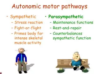
Upper MotorNeurons • Project toLMN • Innervate α and γ motor neurons and inhibitoryinterneurons • Location of UMN • Reticular formation: reticulospinaltract • Vestibular nuclei: vestibulospinaltract • Superior colliculus: tectospinaltract • Red nucleus: rubrospinaltract • Motor cortex: corticospinal & corticobulbar tracts (+ corticorubral & corticoreticular)
DescendingTracts Classification • Classicalclassification: • Pyramidal system • Axons traverse the pyramids inmedulla • Corticospinalaxons • Extrapyramidal system • Other descendingtracts • Basal ganglia and their connections with motorregion • Functionalclassification: • Medialsystem • Innervate medial motornucleus • Lateral system • Innervate lateral motornucleus
DescendingTracts • Lateralpathways • Terminate laterally in the ventral horn • Crossed • Involved in movements of the distal limbs (initiation & finemovement) • Damage – weakness orparalysis • Contains • Lateral corticospinaltract • Ruprospinaltract
Ruprospinaltract • From red nucleus • Crossedimmediately
The Pyramidal (Corticospinal & corticobulbar)Tract • Origin – cerebral cortex • ⅓ from primary motorcortex • ⅓ from premotorareas • ⅓ from primary somatosensorycortex • Terminate in dorsal horn andbrainstem • Modify processing in the somatosensory pathways
CorticospinalTract • Axons passthrough: • Coronaradiata • Internal capsule (posteriorlimb) • Basis pedunculi –midbrain • Medullarypyramids • Decussation • Crossed – Lateral corticospinal90‐95% • Uncrossed ‐ Anterior corticospinal tract
CorticobulbarTract • Same origin & course as corticospinals • Leave tract in brainstem • Terminate in the cranial nerve nuclei • Bilateral ingeneral • Except to facial nucleus
DescendingTracts • Medialpathways • Terminate Medially in the ventralhorn • Trunk & proximal limbmovements • Important in maintaining balance &position • Mostly happenedunconsciously • Damage • Deficits in maintaining balance &posture • Changes in muscletone • Contains: • Reticulospinaltract • Vestibuspinaltract • Tectospinaltract • Anterior corticospinaltract
ReticulospinalTract • From the reticularformation • Important influence on muscle activity andreflexes • Controlled by cortex (corticoreticular) • Contains descending autonomicfibers • – controlled bythalamus • Crossed anduncrossed • Twotracts • Lateral reticulospinal tract – fromMedulla • Medial reticulospinal tract – fromPons
VestibulospinalTract • Importance in maintaining balance • Influence axialmuscles • Uncrossed • Medial & lateral vestibulospinaltracts
TectospinalTract • Superiorcolliculus • Terminate in the cervicalregions • Head movements in response to visual stimuli • Mostly crossed
Anterior CorticospinalTract • Similar to other medialpathways • Terminate in the medial motorn. • Except it is voluntary • Do not cross in pyramidal decussation • May cross beforetermination
Lower Motor NeuronLesion • Flaccid paralysis or paresis(weakness) • Hypo‐ orareflexia • Decreased muscle tone • Atrophy‐ musclewasting • Develops over time(weeks) • Fasciculations – small twitches that are visible to the eye
Upper Motor Neuron Lesion • Paralysis orparesis • Spasticity • Hypertonia • Hyperreflexia • Andmaybe: • Babinskisign • Clonus • Decreased superficialreflexes • Abdominal reflex & Cremastericreflex
Upper Motor Neuron Lesion Babinski Sign • Abnormal response to stroking the lateral planter surface of the foot • Not useful inbabies • Normal response: toesplanter‐flex • Abnormal: dorsiflexion of bigtoe
Upper Motor Neuron Lesion Clonus • Repetitive flexion‐extension of a joint in response to single flexion or extension • https://youtu.be/UX75k8s5QUE
SuperficialReflexes • Decrease With UMNlesions • Abdominal reflex; abdominal muscles contract on stroking the abdomen • Cremasteric reflex (useful in babies); testes elevation with stroking inside of thethigh
Lower-versus Upper-Motor- NeuronLesions Variable Lower-Motor-NeuronLesion Upper-Motor-NeuronLesion Weakness Flaccidparalysis Spasticparalysis Deep tendonreflexes Decreased orabsent Increased Babinski'sreflex Absent Present Atrophy May bemarked Absent or resulting fromdisuse Fasciculations andfibrillations May bepresent Absent
SpinalShock • Follows severe acute injury to the spinalcord • For short period (days orweeks) • Loss of all functions (motor & sensory) bellow level of injury • Loss ofreflexes • Due to sudden loss of supraspinalinputs
Upper MotorNeurons Cerebral Cortex Lateral system Medialsystem Motor Spinalcord & Brainstem cortex Rednu. Retic. Form. Sup. Colliculus α Vestibular Nu. γ Intrafusal Extrafusal Muscle
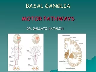
BasalGanglia • The basal ganglia include the caudate, putamen, and globus pallidus and number of closely related nuclei • They influence motor system primarily through projections to upper motorneurons • Motor deficits depend on the specific nucleusdamaged • Understanding the neurochemistry of basal ganglia drives the development of clinicaltreatment
BasalGanglia • The basal ganglia actas • Brake against involuntarymovement • Switch to turn on a fixed actionpattern • Their major output is to the VA of thethalamus • Projects primarily to area 6 (premotor & supplementary motorareas)
Basal GangliaTerminology • Striatum (neostriatum) = caudate +putamen • Lentiform nucleus = putamen + globuspallidus • Corpus striatum = caudate +lentiform • Basal ganglia = corpus striatum +amygdala • Globus pallidus = pallidum =paleostriatum • Claustrum is some times included with the basalganglia • Basal ganglia is included by the extrapyramidalsystem
Basal Ganglia: GrossAnatomy • Caudate nucleus • Parts • Location • Relations • Lateralventricle • Amygdaloidnucleus
Basal Ganglia: GrossAnatomy • Lentiformnucleus • Parts • Putamen • Globus pallidus • Internal(GPi) • External(GPe) • Shape • Location • Relations • External & internal capsules • Claustrum
Basal Ganglia: GrossAnatomy • Amygdaloidnucleus • Subthalamicnucleus • Substantia nigra • Pars reticulata(SNr) • Pars compacta(SNc) • Claustrum
Basal GangliaCircuitry • Inputs • Most inputs enter thestriatum • From cerebral cortex &thalamus • These inputs areexcitatory • Outputs • Most leave from Gpi & SNr • Most go to VA nucleus of the thalamus, which projects to motorcortex • The outputs are GABAergic and inhibitory • VA excites motor cortex, leading to movements • Increase basal ganglia output will inhibit the VA and reduce overallmovements
Basal Ganglia Circuitry Intrinsic Circuits • Large number of connections between components of the basal ganglia • Can be groupedinto • Directpathway • Indirect pathway • These pathways affect the VA activity and thus the motor cortex activity
The DirectPathway • From striatum toGpi • Uses GABA, which inhibits another GABAergic projection (Gpi toVA) • Disinhibition • Cortical activity → ↑direct pathway → ↓Gpi activity → ↑ VAactivity • Activity in the direct pathway leads to increased motor cortex activity and increased movements
The IndirectPathway • Goes from striatum to GPe (GABA) to the subthalamic nucleus (GABA) • Subthalamic nucleus to Gpi (Glu) • ↑ activity in the cortex → ↑ activity of subthalamic nu.→ • ↑ GPi → ↓ VA activity → ↓ motor cortexactivity
Basal GangliaCircuitry • The direct pathway increasemovements • The indirect pathway decrease movements • Normal behavior requires a balance between the direct and indirectpathways • All pathways areuncrossed • Right basal ganglia modulate right cortex and affect movements on the left side of thebody • Acetylcholine is used by the interneurons in the striatum • It affect the output of the direct and indirectpathways • It’s a target for drugtherapy
NigrostriatalPathway • In the striatum different cell typesgive • rise to the direct and indirect pathways • Both cell types receive dopaminergic input from SN parscompacta • These cells have different receptors forDA • For direct pathway, DA excites the striatalcells • For indirect pathway, DA inhibits the striatalcells • Thus the nigrostriatal pathway ↑ the activity of the VA and motorcortex • PD leadsto • ↓ direct pathway activity • ↑ indirect pathway activity • ↓ activity of VA and motorcortex
Cerebellum • The cerebellum is essential for normalmovements • It affects motor behavior by affectingUMNs • The cerebellum acts as acomparator • Compares intended movements (data from cerebral cortex) to the actual movements (sensorydata) • Sends corrective signals into the descending motor pathways
CerebellarFunction • It affects all movements, it is importantin: • Balance • Locomotion • Simple & complexmovements • Eye movements, etc. • Site of motorlearning • Important for learning new motor skills and adjusting movements to changing sensory inputs
Cerebellar Anatomy GrossAnatomy • Location …. • Relations ….. • The cerebellum consists of two hemispheres • The hemispheres are connected by vermis
Cerebellum: GrossAnatomy • Three mainlobes • Anteriorlobe • Primaryfissure • Posterior lobe (middlelobe) • Cerebellartonsils • Posterolateral fissure (uvulonodularfissure) • Flocculonodularlobe
Cerebellum: InternalStructure • Content • Cerebellar cortex (folia) & central nuclei are grey matter • Arbor vitae = tree of life = whitematter
CerebellarAnatomy • Cerebellum includes a cortex & deep nuclei • The deep nuclei are the major source of output from the cerebellum • Four nuclei from medial to lateral • Fastigial • Globose Interposednuclei • Emboliform • Dentate
CerebellarCortex • Cerebellar cortex includes 5 cell types in 3 layers • Five celltypes • Inhibitorycells • Purkinje, basket, Golgi, and stellate cells • Excitatorycells • Granulecells • Three layers • Molecularlayer • Purkinje cell layer • Granule celllayer