Download
1 / 63
630 likes | 663 Views
Learn about the types of bone, bone composition, cellular components, and mechanisms of bone formation to understand the intricate process of bone healing. Explore how osteoblasts, osteocytes, and osteoclasts work together, the role of blood supply in bone healing, and the importance of mechanical stability during fracture repair.

E N D
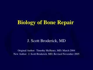
Biology of Bone Repair J. Scott Broderick, MD Original Author: Timothy McHenry, MD; March 2004 New Author: J. Scott Broderick, MD; Revised November 2005
Types of Bone • Lamellar Bone • Collagen fibers arranged in parallel layers • Normal adult bone • Woven Bone (non-lamellar) • Randomly oriented collagen fibers • In adults, seen at sites of fracture healing, tendon or ligament attachment and in pathological conditions
Lamellar Bone • Cortical bone • Comprised of osteons (Haversian systems) • Osteons communicate with medullary cavity by Volkmann’s canals Picture courtesy Gwen Childs, PhD.
Haversian System osteon osteocyte Picture courtesy Gwen Childs, PhD. Haversian canal Volkmann’s canal
Lamellar Bone • Cancellous bone (trabecular or spongy bone) • Bony struts (trabeculae) that are oriented in direction of the greatest stress
Woven Bone • Coarse with random orientation • Weaker than lamellar bone • Normally remodeled to lamellar bone Figure from Rockwood and Green’s: Fractures in Adults, 4th ed
Bone Composition • Cells • Osteocytes • Osteoblasts • Osteoclasts • Extracellular Matrix • Organic (35%) • Collagen (type I) 90% • Osteocalcin, osteonectin, proteoglycans, glycosaminoglycans, lipids (ground substance) • Inorganic (65%) • Primarily hydroxyapatite Ca5(PO4)3(OH)2
Osteoblasts • Derived from mesenchymal stem cells • Line the surface of the bone and produce osteoid • Immediate precursor is fibroblast-like preosteoblasts Picture courtesy Gwen Childs, PhD.
Osteocytes • Osteoblasts surrounded by bone matrix • trapped in lacunae • Function poorly understood • regulating bone metabolism in response to stress and strain Picture courtesy Gwen Childs, PhD.
Osteocyte Network • Osteocyte lacunae are connected by canaliculi • Osteocytes are interconnected by long cell processes that project through the canaliculi • Preosteoblasts also have connections via canaliculi with the osteocytes • Network probably facilitates response of bone to mechanical and chemical factors
Osteoclasts • Derived from hematopoietic stem cells (monocyte precursor cells) • Multinucleated cells whose function is bone resorption • Reside in bone resorption pits (Howship’s lacunae) • Parathyroid hormone stimulates receptors on osteoblasts that activate osteoclastic bone resorption Picture courtesy Gwen Childs, PhD.
Components of Bone Formation • Cortex • Periosteum • Bone marrow • Soft tissue
Prerequisites for Bone Healing • Adequate blood supply • Adequate mechanical stability
Mechanisms of Bone Formation • Cutting Cones • Intramembranous Bone Formation • Endochondral Bone Formation
Cutting Cones • Primarily a mechanism to remodel bone • Osteoclasts at the front of the cutting cone remove bone • Trailing osteoblasts lay down new bone Courtesy Drs. Charles Schwab and Bruce Martin
Intramembranous (Periosteal) Bone Formation • Mechanism by which a long bone grows in width • Osteoblasts differentiate directly from preosteoblasts and lay down seams of osteoid • Does NOT involve cartilage anlage
Intramembranous Bone Formation Picture courtesy Gwen Childs, PhD.
Endochondral Bone Formation • Mechanism by which a long bone grows in length • Osteoblasts line a cartilage precursor • The chondrocytes hypertrophy, degenerate and calcify (area of low oxygen tension) • Vascular invasion of the cartilage occurs followed by ossification (increasing oxygen tension)
Endochondral Bone Formation Picture courtesy Gwen Childs, PhD.
Blood Supply • Long bones have three blood supplies • Nutrient artery (intramedullary) • Periosteal vessels • Metaphyseal vessels Periosteal vessels Nutrient artery Metaphyseal vessels Figure adapted from Rockwood and Green, 5th Ed
Nutrient Artery • Normally the major blood supply for the diaphyseal cortex (80 to 85%) • Enters the long bone via a nutrient foramen • Forms medullary arteries up and down the bone
Periosteal Vessels • Arise from the capillary-rich periosteum • Supply outer 15 to 20% of cortex normally • Capable of supplying a much greater proportion of the cortex in the event of injury to the medullary blood supply
Metaphyseal Vessels • Arise from periarticular vessels • Penetrate the thin cortex in the metaphyseal region and anastomose with the medullary blood supply
Vascular Response in Fracture Repair • Fracture stimulates the release of growth factors that promote angiogenesis and vasodilation • Blood flow is increased substantially to the fracture site • Peaks at two weeks after fracture
Mechanical Stability • Early stability promotes revascularization • After first month, loading and interfragmentary motion promotes greater callus formation
Mechanical Stability • Mechanical load and small displacements at the fracture site stimulate healing • Inadequate stabilization may result in excessive deformation at the fracture site interrupting tissue differentiation to bone (soft callus) • Over-stabilization, however, reduces periosteal bone formation (hard callus)
Stages of Fracture Healing • Inflammation • Repair • Remodeling
Inflammation • Tissue disruption results in hematoma at the fracture site • Local vessels thrombose causing bony necrosis at the edges of the fracture • Increased capillary permeability results in a local inflammatory milieu • Osteoinductive growth factors stimulate the proliferation and differentiation of mesenchymal stem cells
Repair • Periosteal callus forms along the periphery of the fracture site • Intramembranous ossification initiated by preosteoblasts • Intramedullary callus forms in the center of the fracture site • Endochondral ossification at the site of the fracture hematoma • Chemical and mechanical factors stimulate callus formation and mineralization
Repair Figure from Brighton, et al, JBJS-A, 1991.
Remodeling • Woven bone is gradually converted to lamellar bone • Medullary cavity is reconstituted • Bone is restructured in response to stress and strain (Wolff’s Law)
Mechanisms for Bone Healing • Direct (primary) bone healing • Indirect (secondary) bone healing
Direct Bone Healing • Mechanism of bone healing seen when there is no motion at the fracture site (i.e. absolute stability) • Does not involve formation of fracture callus • Osteoblasts originate from endothelial and perivascular cells
Direct Bone Healing • A cutting cone is formed that crosses the fracture site • Osteoblasts lay down lamellar bone behind the osteoclasts forming a secondary osteon • Gradually the fracture is healed by the formation of numerous secondary osteons • A slow process – months to years
Components of Direct Bone Healing • Contact Healing • Direct contact between the fracture ends allows healing to be with lamellar bone immediately • Gap Healing • Gaps less than 200-500 microns are primarily filled with woven bone that is subsequently remodeled into lamellar bone • Larger gaps are healed by indirect bone healing (partially filled with fibrous tissue that undergoes secondary ossification)
Direct Bone Healing Figure from http://www.vetmed.ufl.edu/sacs/notes
Indirect Bone Healing • Mechanism for healing in fractures that have some motion, but not enough to disrupt the healing process. • Bridging periosteal (soft) callus and medullary (hard) callus re-establish structural continuity • Callus subsequently undergoes endochondral ossification • Process fairly rapid - weeks
Local Regulation of Bone Healing • Growth factors • Cytokines • Prostaglandins/Leukotrienes • Hormones • Growth factor antagonists
Growth Factors • Transforming growth factor • Bone morphogenetic proteins • Fibroblast growth factors • Platelet-derived growth factors • Insulin-like growth factors
Transforming Growth Factor • Super-family of growth factors (~34 members) • Acts on serine/threonine kinase cell wall receptors • Promotes proliferation and differentiation of mesenchymal precursors for osteoblasts, osteoclasts and chondrocytes • Stimulates both endochondral and ntramembranous bone formation • Induces synthesis of cartilage-specific proteoglycans and type II collagen • Stimulates collagen synthesis by osteoblasts
Bone Morphogenetic Proteins • Osteoinductive proteins initially isolated from demineralized bone matrix • Proven by bone formation in heterotopic muscle pouch • Induce cell differentiation • BMP-3 (osteogenin) is an extremely potent inducer of mesenchymal tissue differentiation into bone • Promote endochondral ossification • BMP-2 and BMP-7 induce endochondral bone formation in segmental defects • Regulate extracellular matrix production • BMP-1 is an enzyme that cleaves the carboxy termini of procollagens I, II and III
Bone Morphogenetic Proteins • These are included in the TGF-β family • Except BMP-1 • Sixteen different BMP’s have been identified • BMP2-7,9 are osteoinductive • BMP2,6, & 9 may be the most potent in osteoblastic differentiation • Involved in progenitor cell transformation to pre-osteoblasts • Work through the intracellular Smad pathway • Follow a dose/response ratio
Timing and Function of Growth Factors Table from Dimitriou, et al., Injury, 2005
Clinical Use of BMP’s • Used at doses between 10x & 1000x native levels • Negligible risk of excessive bone formation • rhBMP-2 used in “fresh” open fractures to enhance healing and reduce need for secondary procedures after unreamed IM nailing • BESTT study also had a lower infection rate in Type IIIA & B open fractures with application of rhBMP-2 • BMP-7 approved for use in recalcitrant nonunions in patients for whom autografting is not a good option (i.e. medically unstable, previous harvesting of all iliac crest sites, etc.)
BMP Future Directions • BMP-2 • Increased fusion rate in spinal fusion • BMP-7 equally effective as ICBG in nonunions (small series: need larger studies) • Must be applied locally because of rapid systemic clearance • ? Effectiveness in acute fractures • ? Increased wound healing in open injuries • Protein therapy vs. gene therapy • Credibility of researchers compromised
BMP Antagonists • May have important role in bone formation • Noggin • Extra-cellular inhibitor • Competes with BMP-2 for receptors • BMP-13 found to limit differentiation of mesenchymal stromal cells • Inhibits osteogenic differentiation
Fibroblast Growth Factors • Both acidic (FGF-1) and basic (FGF-2) forms • Increase proliferation of chondrocytes and osteoblasts • Enhance callus formation • FGF-2 stimulates angiogenesis
Platelet-Derived Growth Factor • A dimer of the products of two genes, PDGF-A and PDGF-B • PDGF-BB and PDGF-AB are the predominant forms found in the circulation • Stimulates bone cell growth • Mitogen for cells of mesenchymal origin • Increases type I collagen synthesis by increasing the number of osteoblasts • PDGF-BB stimulates bone resorption by increasing the number of osteoclasts
Insulin-like Growth Factor • Two types: IGF-I and IGF-II • Synthesized by multiple tissues • IGF-I production in the liver is stimulated by Growth Hormone • Stimulates bone collagen and matrix synthesis • Stimulates replication of osteoblasts • Inhibits bone collagen degradation
Cytokines • Interleukin-1,-4,-6,-11, macrophage and granulocyte/macrophage (GM) colony-stimulating factors (CSFs) and Tumor Necrosis Factor • Stimulate bone resorption • IL-1 is the most potent • IL-1 and IL-6 synthesis is decreased by estrogen • May be mechanism for post-menopausal bone resorption • Peak during 1st 24 hours then again during remodeling • Regulate endochondral bone formation