Download
1 / 87
900 likes | 1.41k Views
Electron Transport Chain. Thermodynamics of Glucose Oxidation. Glucose + 6 O 2 ——> 6 CO 2 + 6 H 2 O. ∆G o’ = -2866 kJ/mol. Half-Reactions of Glucose Oxidation. Glucose + 6 H 2 O ——> 6 CO 2 + 24 H + + 24 e – 6 O 2 + 24 H + + 24 e – ——> 12 H 2 O. NADH and FADH 2.

E N D
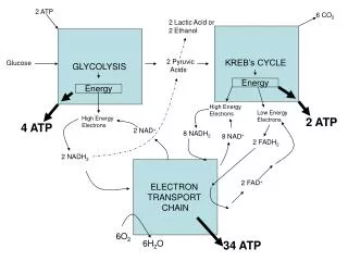
Thermodynamics of Glucose Oxidation Glucose + 6 O2 ——> 6 CO2 + 6 H2O ∆Go’ = -2866 kJ/mol
Half-Reactions of Glucose Oxidation Glucose + 6 H2O ——> 6 CO2 + 24 H+ + 24 e– 6 O2 + 24 H+ + 24 e– ——> 12 H2O NADH and FADH2
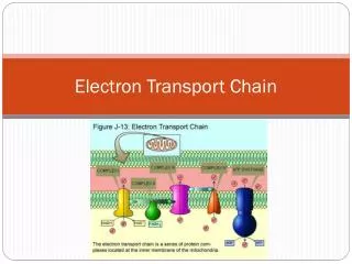
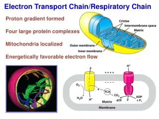
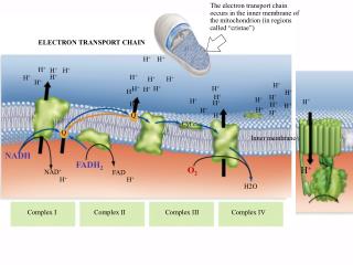
Mitochondrial Electron Transport Chain System of Linked Electron Carriers
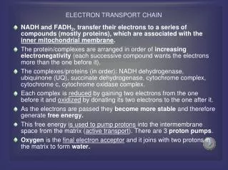
Components of Electron Transport Process • Reoxidation of NADH and FADH2 • Sequential oxidation-reduction of multiple redox centers (four enzyme complexes) • Production of proton gradient across the mitochondrial membrane
Oxidative Phosphorylation Synthesis of ATP driven by free energy of electrochemical gradient
Coupling of Electron Transport and ATP Synthesis NOTE: ATP Synthesis in the Mitochondrion
The Mitochondrion • Prokaryotic origin • Double membrane bound • Genome • Human: encodes 13 genes, all ETC subunits.
Mitochondrial Outer Membrane Permeable to molecules smaller than ~5 kD
X-Ray Structure of E. coli OmpF Porin Figure 9-23a
X-Ray Structure of E. coli OmpF Porin Trimer Figure 9-23b
Mitochondrial Intermembrane Space (IMS)[Metabolites] = Cytosolic ConcentrationLocalized Compartmentation of H+
Types of Transport • Nonmediated Transport (Diffusion) • H2O; O2; CO2 • Mediated Transport • Passive-mediated Transport(facilitated diffusion) • Active Transport • Facilitated by Proteins: • Carriers, Transporters, Translocases, or Permeases.
Kinetic Properties of Mediated Transport • Saturation kinetics • Speed and specificity • Susceptibility to competitive inhibition • Susceptibility to chemical inactivation
Entry of “NADH” into Mitochondria No NADH Transporter
ADP-ATP Translocator ADP/ATP Exchanger Driven by electrochemical gradient
Phosphate Transport Driven by ∆pH
Phosphate Transporter H+(out) H2PO4–(out) H+(in) H2PO4–(in)
Electron Transport Electron Transport is an Exergonic Process
Standard Reduction Potential Difference ∆Eo’ = Eo’(e–acceptor)– Eo’(e–donor) ∆Go’ = –nF∆Eo’ For negative G need positive E E(acceptor) > E(donor) Note: reduction potential is extremely pH sensitive E = Eo’ + 0.06V*(7-pH)
Electron Transport Complexes • Complex I: NADH–Coenzyme Q Oxidoreductase • Complex II: Succinate–Coenzyme Q Oxidoreductase • Complex III: Coenzyme Q–Cytochrome c Oxidoreductase • Complex IV: Cytochrome c Oxidase
Mobile Electron Carriers Coenzyme Q Cytochrome c
Cytochromes Electron Transport Heme Proteins Fe3+ + e– ——> Fe2+
b c a Hemes Note: isoprene side chain Iron-Protoporphyrin IX Like Mb and Hb Note: Thioether Links
Complex I(NADH–Coenzyme Q Oxidoreductase) Accepts Electrons from NADH NADH + CoQ(oxidized) ——> NAD+ + CoQ(reduced) Protons translocated 4H+(Matrix) ——> 4H+(IMS)
Coenzymes of Complex I(Flavin Mononucleotide, FMN)Oxidation states like FAD
Coenzymes of Complex I(Iron-Sulfur Clusters) One-electron oxidation-reduction Conjugated System (Fe between +2 and +3)
Hydrophilic Domain of Complex I from Thermus thermophilis ~ matrix ~ cytoplasm Electrons follow a multistep path
Structure of Bacteriorhodopsin Figure 9-22
Proton Wire • Deprotonation of Schiff base and protonation of Asp 85 • Proton release to the extracellular surface • Reprotonation of the Schiff base and deprotonation of Asp 96 • Reprotonation of Asp 96 from the cytoplasmic surface • Deprotonation of Asp 85 and reprotonation of the proton release site
Complex II(Succinate–Coenzyme Q Oxidoreductase) Contributes Electrons to Coenzyme Q Succinate + CoQ(oxidized) ——> Fumarate + CoQ(reduced) Does not pump protons
Composition of Complex II • Succinate Dehydrogenase • FAD • [4Fe-4S] cluster • [3Fe-4S] cluster • [2Fe-2S] cluster • Cytochrome b560
E. coli Complex II Cytoplasm ~matrix Plasma Membrane ~IM Periplasm ~cytoplasm