Download
1 / 43
470 likes | 958 Views
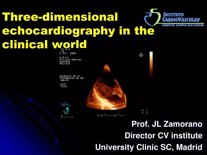
Three-dimensional echocardiography in the clinical world. Prof. JL Zamorano Director CV institute University Clinic SC, Madrid. Advantages of 3D. ... additional advantages ?. B. A. How do you prefer to see a mitral prolapse?. Your preference. B. A. Modes of 3D-Echocardiography.

E N D
Three-dimensional echocardiography in the clinical world Prof. JL Zamorano Director CV institute UniversityClinic SC, Madrid
Advantages of 3D. • ... additional advantages ?. B A How do you prefer to see a mitral prolapse?
Your preference B A
Modes of 3D-Echocardiography. • Full volume. Cropping: every orientation of the planes is possible
Advantages of 3D. • Spatial manipulation. • Optimal alineation of structures. • Views and planes impossible to get in 2D. • Single acquisition, multiple information. • Easy approach to complex problems. • Volumes calculation. • No geometrical assumption (Right ventricle). • Precision MRI, (but faster and cheaper).
Evaluation of Mitral valve area • MVA estimated by: • 2D Echo : • PHT • Planimetry • PISA • 3D • Gorlin • Conclusions: • 3D RT is very accurate in assessing MVA • 3D RT showed better agreement.
MV pre – post MVP Conclusion: 3D RT better correlation with Gorlin, after MVP
Mitral Valve. • Mitral stenosis.
Mitral valve. 3D: Best diagnostic tool for structural mitral evaluation. • Mitral stenosis. • Anatomy of leaflets, comissures, subvalvular apparatus. • Planimetry of oriffice: superior to 2D. • Guide for balloon valvulotomy. • Score. • Oriffice post-valvuloplastia. • Complications.
Circulation, March 2003 Mitral valve. • Mitral regurgitation. • Mechanism of Mi regurgitation
Mitral valve. • Mitral regurgitation. • Volumes quantification. • 3D colour doppler quantification: promising perspectives. • Mechanism of Mi regurgitation.
A3 P3 A2 P2 A1 P1 3D MVP. • 3D Protocol: 4 steps • 1) P short axis + cutting planes
3D MV Prolapse • 3D Protocol: 4 steps • 1) P Shurt axis guided cutting planes • 2) Apical + guided planes • 3) Full volume • 4) 3D color. MI.
1.- 100% P2 2.- False negative (anterolateral region) 3.- False positive (inferomedial region) P2 A1-P1 A3-P3 Agreement 3D vs TEE p< 0,0001
2D: Simpson Problems: ¿Optimal alignement ? Geometric asumptions Volumes. 2D echo ??
3D underestimates volumes • 3D: Volumes calculation Quantification of left ventricular volumes and ejection fraction using freehand transthoracic three-dimensional echocardiography: comparison with magnetic resonance imaging. Mannaerts HF, Van Der Heide JA, Kamp O, Papavassiliu T, Marcus JT, Beek A, Van Rossum AC, Twisk J, Visser CA.JASE 2003
3D. Volumes • 2D: Simpson biplane • 3D (FV): • 2 planes • 4 planes • 8 planes • CMR: Simpson
RT3D: Semiautomated border detection Improved semiautomated quantification of LV volumes and EF using 3D echocardiography with a full matrix-array transducer: comparison with magnetic resonance imaging. Caiani EG, Corsi C, Zamorano JL, Sugeng L, MacEneaney P, Weinert L, Battani R, Gutiérrez-Chico JL, Koch R, Pérez de Isla L, Mor-Avi V, Lang RM.JASE 2005 Aug
Volúmenes 3D • Volumes calculation – Slice view • Direct comparison with gold standard
DTSVI IVTTSVI AAo = π ( )2 2IVTAo Aortic stenosis: Continuity equation • Cumulative error in several parameters • Depending on good parasternal & apical acoustic windows TSVI IVT TSVI
DTSVI IVTTSVI AAo = π ( )2 2IVTAo Aortic stenosis: Continuity equation • Cumulative error in several parameters • Depending on good parasternal & apical acoustic windows TSVI IVT TSVI STROKE VOLUME
SV AAo = IVTAo Aortic stenosis: Continuity equation • Cumulative error in several parameters • Depending on good parasternal & apical acoustic windows TSVI IVT TSVI STROKE VOLUME
Aortic area: • Invasive: • Gorlin • Hakki • Echo: • Continuity equation • Volumetric Simpson • RT 3D
Aortic area with RT3D: Results • Correlation (linear association): Pearson r • Absolute agreement: • ICCa (Intraclass correlation coefficient – absolute) • Lin’s coefficient
Limitations of 3D. • Quantification off-line ??. • Time-consuming. • Full-volume mode: breath-hold. • Image definition does not improve respect to 2D. • Lower frame-rate.
Conclusions • 3D is the best imaging for Mitral valve. • New approach to mitral anatomy and mitral prolapse. • 3D planimetry:Best non-invasive methode for Mi stenosis. • Mechanism of Mi regurgitation. • Best option for complex problems, MVP, surgical repair. • 3D colour Doppler: promising perspectives for quantification of valvular regurgitations. • Volume estimation… similar to MRI
Exécutif Législatif 3D !!! Judiciaire 3 is the number !