Download
1 / 11
490 likes | 1.9k Views
Tumor Necrosis Factor-Alpha (TNF-α) A Cytokine Protein Involved In Inflammatory and Immunologic Responses By: Mourad Ali. Background and Nomenclature.

E N D
Tumor Necrosis Factor-Alpha (TNF-α)A Cytokine Protein Involved In Inflammatory and Immunologic ResponsesBy: Mourad Ali Mourad Ali BCMB 8010, Fall 2008
Background and Nomenclature • Cytokines (from the Greek "cyto" means cell, and "kinos" means movement) are a family of proteins and glycoproteins that are used in cell signaling. Their production is usually triggered by infection, inflammation, or cancer. • Tumor Necrosis Factors (TNF) represent a subfamily of the cytokines. TNF family include the two members Tumor Necrosis Factor-alpha (TNF-α) and Tumor Necrosis Factor-beta (TNF-β). • Macrophages were found to secrete a factor that was able to kill mouse fibrosarcoma and it was named Tumor Necrosis Factor. Mourad Ali BCMB 8010, Fall 2008
Ribbon diagrams display of TNF- α homotrimer. The figure shows the three monomers that form TNF- α. Crystal structure is taken from PDB file 1tnf. The structure is taken from the following article: Eck, M. J., and Sprang, S. R. (1989) J Biol Chem 264, 17595-17605 Mourad Ali BCMB 8010, Fall 2008
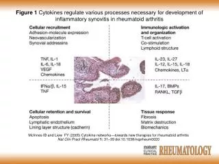
Biological Roles of TNF-α Some of the biological roles played by TNF-α include: • Kills tumor cells. • Regulates the adherence of neutrophils to the endothelium. • Stimulates of the immune system and combating infectious agents. • Regulates sleeping pattern. • Controls embryogenesis. • Modulates the release of corticotrophin from the pituitary gland. Mourad Ali BCMB 8010, Fall 2008
TNF/Receptor Interactions Mourad Ali BCMB 8010, Fall 2008
Ribbon diagram display of TNF- β monomer bound to the soluble extracellular domain of TNF-R55 receptor. Crystal structure is taken from PDB file 1tnr. The structure is taken from the following article: Banner, D. W., D'Arcy, A., Janes, W., Gentz, R., Schoenfeld, H. J., Broger, C., Loetscher, H., and Lesslauer, W. (1993) Cell 73, 431-445 Mourad Ali BCMB 8010, Fall 2008
Diseases with High TNF- α Levels • Several auto-immune diseases are associated with the presence of high activity of TNF-α which induces strong inflammatory response in tissues. • Examples of such diseases are: Rheumatoid arthritis, ankylosing spondylitis, Crohn’s disease and psoriasis Mourad Ali BCMB 8010, Fall 2008
Inhibition of TNF- α • Monoclonal antibodies can be used to treat inflammatory diseases which are characterized by the presence of high levels of TNF- α. Example of a monoclonal antibody that specifically binds and inhibits TNF- α. is Infliximab (Remicade). • Another way to reduce the elevated levels of TNF- α is the use of circulating receptor fusion protein such as Etanercept (Enbrel). Mourad Ali BCMB 8010, Fall 2008
Small-Molecule Inhibitor of TNF- α • This molecule inhibits TNF- α activity through accelerating the dissociation of the TNF- α trimer. It displaces one subunit of the TNF- α trimer and forms an intermediate complex with the remaining two subunits of TNF- α. The dissociation rate is 600-fold greater with the intermediate complex than the un-complexed TNF- α trimer. • There are 16 amino acid residues on the TNF- α dimer that contact the inhibitor, nine of these residues are on chain A and seven are on chain B. although the inhibitor is buried within the TNF- α dimer but it does not show any hydrogen bonds or salt bridges interacting the dimer. The inhibitor-dimer interaction is presumed to be hydrophobic. Mourad Ali BCMB 8010, Fall 2008
Ribbon diagram display of small-molecule inhibitor bound to TNF-α dimer. Crystal structure is taken from PDB file 2az5. The structure is taken from the following article: He, M. M., Smith, A. S., Oslob, J. D., Flanagan, W. M., Braisted, A. C., Whitty, A., Cancilla, M. T., Wang, J., Lugovskoy, A. A., Yoburn, J. C., Fung, A. D., Farrington, G., Eldredge, J. K., Day, E. S., Cruz, L. A., Cachero, T. G., Miller, S. K., Friedman, J. E., Choong, I. C., and Cunningham, B. C. (2005) Science 310, 1022-1025 Mourad Ali BCMB 8010, Fall 2008
Ribbon diagram display of small-molecule inhibitor bound to TNF-α. The figure shows the inhibitor molecule and the 16 contact residues on the TNF-α dimer that constitute the binding surface for the inhibitor. The structure is taken from the following article: He, M. M., Smith, A. S., Oslob, J. D., Flanagan, W. M., Braisted, A. C., Whitty, A., Cancilla, M. T., Wang, J., Lugovskoy, A. A., Yoburn, J. C., Fung, A. D., Farrington, G., Eldredge, J. K., Day, E. S., Cruz, L. A., Cachero, T. G., Miller, S. K., Friedman, J. E., Choong, I. C., and Cunningham, B. C. (2005) Science 310, Mourad Ali BCMB 8010, Fall 2008