Download
1 / 5
50 likes | 65 Views
Human IL-12 (p70) ELISA Kit can be provided from Creative Diagnostics.t<br>http://www.creative-diagnostics.com/Human-IL-12-p70-ELISA-Kit-122853-463.htm<br>

E N D
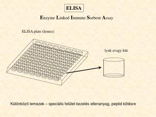
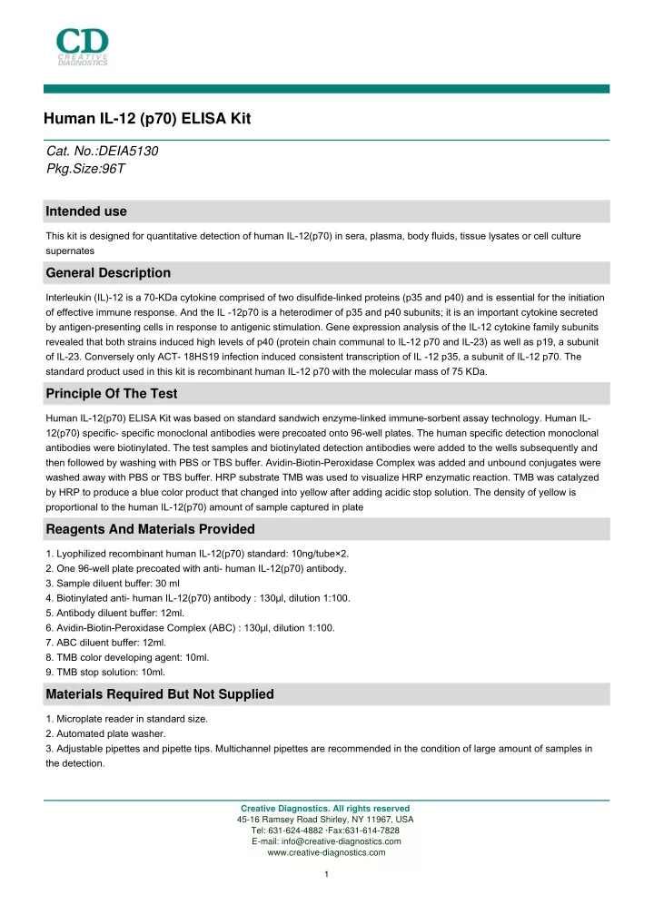
Human IL-12 (p70) ELISA Kit Cat. No.:DEIA5130 Pkg.Size:96T Intended use This kit is designed for quantitative detection of human IL-12(p70) in sera, plasma, body fluids, tissue lysates or cell culture supernates General Description Interleukin (IL)-12 is a 70-KDa cytokine comprised of two disulfide-linked proteins (p35 and p40) and is essential for the initiation of effective immune response. And the IL -12p70 is a heterodimer of p35 and p40 subunits; it is an important cytokine secreted by antigen-presenting cells in response to antigenic stimulation. Gene expression analysis of the IL-12 cytokine family subunits revealed that both strains induced high levels of p40 (protein chain communal to IL-12 p70 and IL-23) as well as p19, a subunit of IL-23. Conversely only ACT- 18HS19 infection induced consistent transcription of IL -12 p35, a subunit of IL-12 p70. The standard product used in this kit is recombinant human IL-12 p70 with the molecular mass of 75 KDa. Principle Of The Test Human IL-12(p70) ELISA Kit was based on standard sandwich enzyme-linked immune-sorbent assay technology. Human IL- 12(p70) specific- specific monoclonal antibodies were precoated onto 96-well plates. The human specific detection monoclonal antibodies were biotinylated. The test samples and biotinylated detection antibodies were added to the wells subsequently and then followed by washing with PBS or TBS buffer. Avidin-Biotin-Peroxidase Complex was added and unbound conjugates were washed away with PBS or TBS buffer. HRP substrate TMB was used to visualize HRP enzymatic reaction. TMB was catalyzed by HRP to produce a blue color product that changed into yellow after adding acidic stop solution. The density of yellow is proportional to the human IL-12(p70) amount of sample captured in plate Reagents And Materials Provided 1. Lyophilized recombinant human IL-12(p70) standard: 10ng/tube×2. 2. One 96-well plate precoated with anti- human IL-12(p70) antibody. 3. Sample diluent buffer: 30 ml 4. Biotinylated anti- human IL-12(p70) antibody : 130μl, dilution 1:100. 5. Antibody diluent buffer: 12ml. 6. Avidin-Biotin-Peroxidase Complex (ABC) : 130μl, dilution 1:100. 7. ABC diluent buffer: 12ml. 8. TMB color developing agent: 10ml. 9. TMB stop solution: 10ml. Materials Required But Not Supplied 1. Microplate reader in standard size. 2. Automated plate washer. 3. Adjustable pipettes and pipette tips. Multichannel pipettes are recommended in the condition of large amount of samples in the detection. Creative Diagnostics. All rights reserved 45-16 Ramsey Road Shirley, NY 11967, USA Tel: 631-624-4882 ·Fax:631-614-7828 E-mail: info@creative-diagnostics.com www.creative-diagnostics.com 1
4. Clean tubes and Eppendorf tubes. 5. Washing buffer (neutral PBS or TBS). Preparation of 0.01M TBS: Add 1.2g Tris, 8.5g Nacl; 450μl of purified acetic acid or 700μl of concentrated hydrochloric acid to 1000ml H2O and adjust pH to 7.2-7.6. Finally, adjust the total volume to 1L. Preparation of 0.01 M PBS: Add 8.5g sodium chloride, 1.4g Na2HPO4 and 0.2g NaH2PO4 to 1000ml distilled water and adjust pH to 7.2-7.6. Finally, adjust the total volume to 1L Storage 4℃ for frequent use -20℃ for infrequent use Avoid multiple freeze-thaw cycles (Shipped with wet ice.) Expiration: Four months at 4℃; Eight months at -20℃ Specimen Collection And Handling 1. Sample Preparation and Storage Store samples to be assayed within 24 hours at 2-8°C. For long-term storage, aliquot and freeze samples at -20°C. Avoid repeated freeze-thaw cycles 1) Cell culture supernate, tissue lysate or body fluids: Remove particulates by centrifugation, analyze immediately or aliquot and store at -20°C 2) Serum: Allow the serum to clot in a serum separator tube (about 30 min) at room temperature. Centrifuge at approximately 1000 X g for 10 min. Analyze the serum immediately or aliquot and store frozen at -70°C 2. Sample Dilution Guideline The user needs to estimate the concentration of the target protein in the sample and select a proper dilution factor so that the diluted target protein concentration falls near the middle of the linear regime in the standard curve. Dilute the sample using the provided diluent buffer. The following is a guideline for sample dilution. Several trials may be necessary in practice. The sample must be well mixed with the diluents buffer 1) High target protein concentration (5-50ng/ml). The working dilution is 1:100. i.e. Add 1 μl sample into 99 μl sample diluent buffer. 2) Medium target protein concentration (500-5000pg/ml). The working dilution is 1:10. i.e. Add 10 μl sample into 90 μl sample diluent buffer. 3) Low target protein concentration (7.8-500pg/ml). The working dilution is 1:2. i.e. Add 50 μl sample to 50 μl sample diluent buffer. 4) Very Low target protein concentration (≤7.8pg/ml). No dilution necessary, or the working dilution is 1:2 Reagent Preparation 1. Reconstitution of the human IL-12(p70) standard: IL-12(p70) standard solution should be prepared no more than 2 hours prior to the experiment. Two tubes of IL-12(p70) standard (10ng per tube) are included in each kit. Use one tube for each experiment. 1) 10,000pg/ml of human IL-12(p70) standard solution: Add 1 ml sample diluent buffer into one tube, keep the tube at room temperature for 10 min and mix thoroughly. 2) 500pg/ml of human IL-12(p70) standard solution: Add 0.05 ml of the above 10ng/ml IL-12(p70) standard solution into 0.95 ml sample diluent buffer and mix thoroughly. 3) 250pg/ml → 7.8pg/ml of human IL-12(p70) standard solutions: Label 6 Eppendorf tubes with 250pg/ml, 125pg/ml, 62.5pg/ml, 31.3pg/ml, 15.6pg/ml, 7.8pg/ml, respectively. Aliquot 0.3 ml of the sample diluent buffer into each tube. Add 0.3 ml of the above 500pg/ml IL-12(p70) standard solution into 1st tube and mix. Transfer 0.3 ml from 1st tube to 2nd tube and mix. Transfer 0.3 ml Creative Diagnostics. All rights reserved 45-16 Ramsey Road Shirley, NY 11967, USA Tel: 631-624-4882 ·Fax:631-614-7828 E-mail: info@creative-diagnostics.com www.creative-diagnostics.com 2
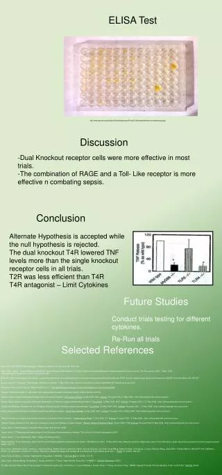
from 2nd tube to 3rd tube and mix, and so on. Note: The standard solutions are best used within 2 hours. The 10ng/ml standard solution may be stored at 4°C for up to 12 hours, or at -20°C for up to 48 hours. Avoid repeated freeze-thaw cycles. 2. Preparation of biotinylated anti-human IL-12(p70) antibody working solution: The solution should be prepared no more than 2 hours prior to the experiment. 1) The total volume should be: 0.1ml/well x the number of wells (allowing 0.1-0.2 ml more than total volume) 2) Biotinylated anti-human IL-12(p70) antibody should be diluted in 1:100 with the antibody diluent buffer and mixed thoroughly. 3. Preparation of Avidin-Biotin-Peroxidase Complex (ABC) working solution: The solution should be prepared no more than 1 hour prior to the experiment. 1) The total volume should be: 0.1ml/well x the number of wells (allowing 0.1-0.2 ml more than total volume) 2) Avidin- Biotin-Peroxidase Complex (ABC) should be diluted in 1:100 with the ABC dilution buffer and mixed thoroughly Assay Steps The ABC working solution and TMB color developing agent must be kept warm at 37°C for 30 min before use. When diluting samples and reagents, they must be mixed completely and evenly. Standard IL-12(p70) detection curve should be prepared for each experiment. The user will decide sample dilution fold by crude estimation of IL-12(p70) amount in samples 1. Aliquot 0.1ml per well of the 500pg/ml, 250pg/ml, 125pg/ml, 62.5pg/ml, 31.3pg/ml, 15.6pg/ml, 7.8pg/ml human IL-12(p70) standard solutions into the precoated 96-well plate. Add 0.1ml of the sample diluent buffer into the control well (Zero well). Add 0.1ml of each properly diluted sample of human sera, plasma, body fluids, tissue lysates or cell culture supernatants to each empty well. We recommend that each human IL-12(p70) standard solution and each sample is measured in duplicate. 2. Seal the plate with the cover and incubate at 37°C for 90 min. 3. Remove the cover, discard plate content, and blot the plate onto paper towels or other absorbent material. Do NOT let the wells completely dry at any time. 4. Add 0.1ml of biotinylated anti-human IL-12(p70) antibody working solution into each well and incubate the plate at 37°C for 60 min. 5. Wash plate 3 times with 0.01M TBS or 0.01M PBS, and each time let washing buffer stay in the wells for 1 min. Discard the washing buffer and blot the plate onto paper towels or other absorbent material. Plate Washing Method: Discard the solution in the plate without touching the side walls. Blot the plate onto paper towels or other absorbent material. Soak each well with at least 0.3 ml PBS or TBS buffer for 1~2 minutes. Repeat this process two additional times for a total of THREE washes. Note: For automated washing, aspirate all wells and wash THREE times with PBS or TBS buffer, overfilling wells with PBS or TBS buffer. Blot the plate onto paper towels or other absorbent material. 6. Add 0.1ml of prepared ABC working solution into each well and incubate the plate at 37°C for 30 min. 7. Wash plate 5 times with 0.01M TBS or 0.01M PBS, and each time let washing buffer stay in the wells for 1-2 min. Discard the washing buffer and blot the plate onto paper towels or other absorbent material. 8. Add 90 μl of prepared TMB color developing agent into each well and incubate plate at 37°C in dark for 15-20 min Note: For reference only, the optimal incubation time should be determined by end user. And the shades of blue can be seen in the wells with the four most concentrated human IL-12(p70) standard solutions; the other wells show no obvious color 9. Add 0.1ml of prepared TMB stop solution into each well. The color changes into yellow immediately. 10. Read the O.D. absorbance at 450nm in a microplate reader within 30 min after adding the stop solution Calculation For calculation, (the relative O.D.450 ) = (the O.D. 450 of each well) – (the O.D.450 of Zero well). The standard curve can be plotted as the relative O.D. 450 of each standard solution (Y) vs. the respective concentration of the standard solution (X). The human IL-12(p70) concentration of the samples can be interpolated from the standard curve. Creative Diagnostics. All rights reserved 45-16 Ramsey Road Shirley, NY 11967, USA Tel: 631-624-4882 ·Fax:631-614-7828 E-mail: info@creative-diagnostics.com www.creative-diagnostics.com 3
Note: If the samples measured were diluted, multiply the dilution factor to the concentrations from interpolation to obtain the concentration before dilution Typical Standard Curve This standard curve was generated for demonstration purpose only. A standard curve must be run with each assay. Detection Range 7.8pg/ml-500pg/ml Sensitivity < 2 pg/ml Specificity No detectable cross-reactivity with any other cytokine Limitations 1. Before using Kit, spin tubes and bring down all components to bottom of tube. 2. Duplicate well assay was recommended for both standard and sample testing. 3. Don’t let 96-well plate dry, dry plate will inactivate active components on plate. 4. In order to avoid marginal effect of plate incubation due to temperature difference ( reaction may be stronger in the marginal wells), it is suggested that the diluted ABC and TMB solution will be pre-warmed in 37 for 30 min before using. REFERENCES 1. Toubai T, Tanaka J, Ota S, Fukuhara T, Hashino S, Kondo T, Shono Y, Morioka M, Kawamura T, Masauzi N, Kakinoki Y, Kobayashi H, Kunieda Y, Kasai M, Kurosawa M, Asaka M, Imamura M. Effect of granulocyte colony-stimulating factor on IL-12 Creative Diagnostics. All rights reserved 45-16 Ramsey Road Shirley, NY 11967, USA Tel: 631-624-4882 ·Fax:631-614-7828 E-mail: info@creative-diagnostics.com www.creative-diagnostics.com 4
p40 production during chemotherapy for B-cell lineage non-Hodgkin's lymphoma patients. Eur J Haematol. Nov;77(5):403- 9.2006. 2. Nakamura T, Kimura H, Kato M, Kurashige S, Wakamatsu K. A sensitive and reliable quantification method for mouse interleukin-12 p70 based on fluorometric sandwich ELISA (FS-ELISA).Cell Biol Int. Feb;31(2):173-9.2007 3. Spensieri F, Fedele G, Fazio C, Nasso M, Stefanelli P, Mastrantonio P, Ausiello CM. Bordetella pertussis inhibition of interleukin-12 (IL-12) p70 in human monocyte-derived dendritic cells blocks IL-12 p35 through adenylate cyclase toxin-dependent cyclic AMP induction. Infect Immun. May;74(5):2831-8,2006. Creative Diagnostics. All rights reserved 45-16 Ramsey Road Shirley, NY 11967, USA Tel: 631-624-4882 ·Fax:631-614-7828 E-mail: info@creative-diagnostics.com www.creative-diagnostics.com 5