Download

1 / 25
660 likes | 2.55k Views
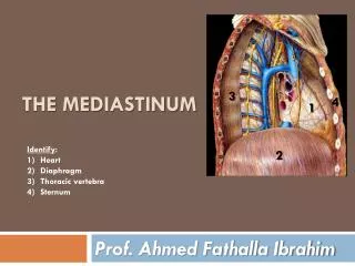
3. 4. 1. Identify : Heart Diaphragm Thoracic vertebra Sternum. 2. THE mediastinum. Dr. Ahmed Fathalla Ibrahim. OBJECTIVES. At the end of the lecture, students should be able to: Define the “ Mediastinum ”. Differentiate between the divisions of the mediastinum .

E N D
3 4 1 Identify: Heart Diaphragm Thoracic vertebra Sternum 2 THE mediastinum Dr. Ahmed Fathalla Ibrahim
OBJECTIVES At the end of the lecture, students should be able to: • Define the “Mediastinum”. • Differentiate between the divisions of the mediastinum. • List the boundaries and contents of each division. • Describe the relations between the important structures in each division.
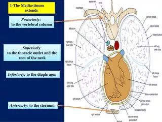
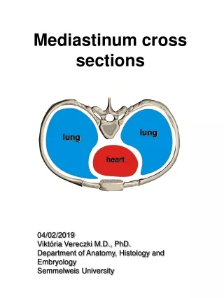
THE MEDIASTINUM • It is a partition between right & left pleural sacs & lungs. • It includes all the structures which lie in the intermediate compartments of the thoracic cavity.
BOUNDARIES OF MEDIASTINUM • Superior: Thoracic outlet • Inferior: Diaphragm • Anterior: Sternum • Posterior: Thoracic vertebrae • Lateral:Lungs & pleurae
DIVISIONS OF THE MEDIASTINUM It is divided by a horizontal plane extending from sternal angle to lower border of 4th thoracic vertebra into: 1. Superior mediastinum (S): above the plane 2. Inferior mediastinum: below the plane, Inferior mediastinum is subdivided into: Middle mediastinum (M): contains heart Anterior mediastinum (A): in front of heart Posterior mediastinum (P): behind heart S A M P
BOUNDARIES OF SUPERIOR MEDIASTINUM • Superior: Thoracic outlet • Inferior: Horizontal plane • Anterior: Manubrium of sternum • Posterior: Upper 4 thoracic vertebrae • Lateral:lungs & pleurae
CONTENTS OF SUPERIOR MEDIASTINUM Thoracic duct Left vagus nerve Trachea Right vagus nerve Left common carotid artery Left subclavian artery Brachiocephalic artery Left brachiocephalic vein Right phrenic nerve Thymus Right brachiocephalic vein Left phrenic nerve Arch of aorta S V C Esophagus
CONTENTS OF SUPERIOR MEDIASTINUM c b a A A B FROM SUPERFICIAL TO DEEP: Thymus gland Veins: -Right & left brachiocephalic -Superior vena cava Arteries: -Arch of aorta (A) & its branches a-Brachiocephalic artery b-Left common carotid c-Left subclavian Nerves: - Right & left vagus -Right & left phrenic 5. Trachea 6. Esophagus 7. Thoracic duct 8. Lymph nodes C
CONTENTS OF SUPERIOR MEDIASTINUM • 4 ARTERIES: arch of aorta, brachiocephalic, left common carotid, left subclavian • 4 NERVES: right & left vagus, right & left phrenic • 3 VEINS: right & left brachiocephalic, SVC • 2 TUBES: trachea & esophagus • 1 GLAND: thymus • 1 DUCT: thoracic duct
BOUNDARIES OF POSTERIOR MEDIASTINUM • Superior: Horizontal plane • Inferior: Diaphragm • Anterior: Heart • Posterior: Thoracic vertebrae from T5 to T12 • Lateral: Lungs & pleurae
CONTENTS OF POSTERIOR MEDIASTINUM Esophagus Vagus nerves: around esophagus Thoracic duct: posterior to esophagus Azygos vein: posterior & to the right of esophagus Descending aorta: posterior & to the left of esophagus Right & left sympathetic trunks Lymph nodes
CONTENTS OF POSTERIOR MEDIASTINUM E S O P H A G U S Right & left Vagus nerves Left Sympathetic Trunk Right Sympathetic Trunk
MIDDLE MEDIASTINUM SITE: • Between anterior & posterior mediastinum CONTENTS: • Heart & pericardium • Ascending Aorta • Pulmonary trunk • Superior & inferior vena cava • Right & left pulmonary veins • Right & left phrenic nerves • Lymph nodes Left Pulmonary veins Right Pulmonary veins Left Phrenic nerve Right Phrenic nerve Heart Diaphragm Inferior vena cava
ANTERIOR MEDIASTINUM BOUNDARIES: • Superior: Horizontal plane • Inferior: Diaphragm • Anterior: Body & xiphoid process of sternum • Posterior: Heart • Lateral: Lungs & pleurae CONTENTS: • Thymus gland • Lymph nodes
LEVEL OF T4 • Level of: • Sternal angle • Second costal cartilage • Level of: • Bifurcation of trachea • Bifurcation of pulmonary • trunk • Beginning & termination of • arch of aorta
VAGUS NERVE Left common carotid • It is the 10th cranial nerve. • The right vagusdescends to the right side of trachea, forms the posterior esophageal plexus & continues in abdomen as posterior gastric nerve. • The left vagusdescends between left common carotid & left subclavian arteries, forms the anterior esophageal plexus & continues in abdomen as anterior gastric nerve. Left subclavian
PHRENIC NERVE ROOT VALUE: • C3,4,5 COURSE IN THORAX: • The right phrenicdescends on the right side of SVC & heart. • The left phrenicdescends on the left side of heart. • Both nerves terminate in the diaphragm • SUPPLY: 1) Motor & sensory fibers to diaphragm 2) Sensory fibers to pleurae & pericardium
LYMPHATIC VESSELS IN THORAX Lymph form right side of head & neck Lymph from left side of head & neck Lymph from right upper limb Lymph from left upper limb end end Lymph from right side of thorax Lymph from left side of thorax Superior Mediastinum E S O P H A G U S Posterior Mediastinum Cysternachyli (contains lymph from lower half of body)
THORACIC DUCT BEGINNING: • It is the continuation of cysternachyli. COURSE: • It passes through aortic opening of diaphragm. • It ascends in posteriormediastinum (posterior to esophagus). • It ascends in superior mediastinum (to the left of esophagus). TRIBUTARIES: It receives: • Lymphatics from all body EXCEPT: right side of thorax, right upper limb & right side of head & neck. END: • It ends in the left brachiocephalic vein.
AORTA • ASCENDING AORTA: • Beginning: at aortic orifice of left ventricle. • Course: in middle mediastinum • End: continues as arch of aorta (at level of T4) • ARCH OF AORTA: • Course: in superior mediastinum • End: continues as descending thoracic aorta (at level of T4) • DESCENDING AORTA: • Course: in posterior mediastinum • End: continues as abdominal aorta through diaphragm Arch Asc. Ao. Descending aorta
QUESTION 1 • Which one of the following structures is present in the superior mediastinum? • Ascending aorta • Arch of aorta • Descending aorta • Pulmonary trunk
QUESTION 2 • Which one of the following structures is present in both superior & middle mediastinum? • Superior vena cava • Pulmonary trunk • Trachea • Descending aorta
QUESTION 3 • Which one of the following structures lies on the left side of esophagus in the posterior mediastinum? • Superior vena cava • Descending aorta • Azygos vein • Pulmonary trunk