Download
1 / 51
540 likes | 573 Views
Explore the anatomy and clinical presentation of mediastinal lesions, along with imaging and diagnostic techniques. Learn about common tumors and cysts, and how to obtain tissue diagnosis for solid masses. Gain insights into specific mediastinal conditions like thymoma and germ cell tumors.

E N D
Lesions of the Mediastinum Julye Carew, M.D. December 10, 2004
Lesions of the Mediastinum • Anatomy of mediastinum • Clinical Presentation of mediastinal disease • Imaging Techniques • Diagnostic Techniques • Tumors and cysts of the mediastinum
Caveat • Most common “mediastinal mass” is involvement by bronchogenic carcinoma • Limit discussion to primary mediastinal lesions
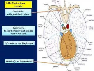
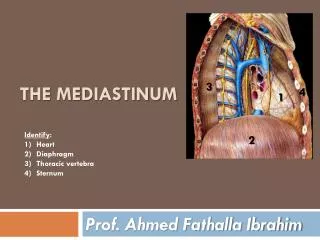
Mediastinal Anatomy • ANTERIOR (includes “superior”) • bordered by sternum and heart • Contains thymus, thyroid, parathyroid, lymphatics
Mediastinal Anatomy • MIDDLE • Anterior border comprised of anterior heart border, and posteriorly by posterior heart border and trachea • Contains heart, trachea, aortic arch, pulmonary arteries, pulmonary hila and lymph nodes
Mediastinal Anatomy • POSTERIOR • Bordered by posterior heart and trachea and vertebrae • Contains esophagus, descending aorta, azygous and hemiazygous veins, paravertebral LN, sympathetic chain and thoracic duct
Mediastinal Anatomy Albert: Clinical Respiratory Medicine, 2nd ed, p.790
Most common presentation- asymptomatic Airway compression with post-obstructive pneumonia Dysphagia Paralysis Hoarseness- RLN Elevated HD- phrenic Horner’s syndrome- sympathetic chain SVC syndrome Hemoptysis, CP CONSTITUTIONAL Clinical Presentation
Imaging- Radiographs • Screening technique • Diagnostic for pneumomediastinum • For all other abnormalities, CT
Computed Tomography • Helpful in determining exact location of mass and density (cystic, fat, vascular, soft tissue) • Always use contrast if possible
MRI • Typically adds little to CT with contrast, except: • 1. Contrast allergies • 2. Multiplanar imaging • 3. Neurogenic tumors • 4. Delineation of vascular invasion • 5. Complex fluid collections • Long data acquisition time/ breathholding
PET scan • Most commonly used as adjunctive mediastinal staging modality in bronchogenic CA (93% sens, 98% spec) • Helpful in clinically staged I and III patients • NOT routinely used to work-up primary mediastinal lesions
Tissue Diagnosis • Solid masses and LN enlargement require biopsy for definitive diagnosis • FNA • Mediastinoscopy • EUS
FNA • Performed via bronchoscopy or by CT-guidance • Bronchoscopic- “blind” with varying degrees of sens/spec • Bronchoscopy only allows for FNA of subcarinal or right paratracheal • Of limited utility in lymphoma, neuroendocrine tumors
FNA by Bronchoscopy Strollo, Chest 1997; 112: 1345
FNA • CT-guidance • PHD- no core biopsies, limiting diagnostic yield for certain tumors • More risky in patients with obstructive lung disease, or functional limitations • A negative or “non-diagnostic” biopsy does not exclude malignant process
Endoscopic Ultrasound • Most commonly used for mediastinal masses adjacent to the esophagus • Larsen et al. looked at 84 patient referred for EUS (all lesions adjacent to esophagus), 34 with known lung primary and 50 with unknown primaries
EUS Larsen et al., 2002 Known Lung Primary
EUS, Larsen, et al., 2002 Unknown Primary
EUS • In patients with lung primary- for nodal evaluation, sens=90%, spec=100%, PPV=100%, NPV=82% • In patients with unknown primary- sens=92%, spec=100%, PPV=100%, NPV=79%
Mediastinoscopy/Thoracotomy • Gold standard • Allows direct visualization of LN, mass in anterior and superior mediastinum, including right paratracheal, left paratracheal to level of aortic arch • Provides larger specimens for histologic examination • Subcarinal and AP lesions require second intercostal space approach
Thymic neoplasms Germ Cell tumors Lymphoma Thyroid neoplasms Parathyroid neoplasms Mesenchymal tumors (lipoma, fibroma, hemangioma, lymphangioma) Primary carcinoma Angiofollicular lymphoid hyperplasia (Castleman’s) Anterior Mediastinum
Thymoma • Most common primary tumor of the anterior mediastinum • Up to half suffer from MG, hypogammaglobulinemia, or pure red cell aplasia • Only 15% of patient with MG have a thymoma- always check Ach receptor antibody levels in diagnosed thymomas
Thymoma Strollo, Chest 1997; 112: 514
Thymoma • Epithelial neoplasms • Most are surrounded by fibrous capsule, but may invade vital structures • Metastasis is rare • Can seed the pleural space but effusion is rare • Goal is complete resection, with XRT for incompletely excised tumors and consideration of cisplatin based chemoTx
Thymic Carcinoma Strollo, Chest 1997; 112: 515
Thymic Carcinoma • Most commonly SCC (differentiate from lung primary) • Aggressive with local invasion and mets • Frequently associated with pleural and pericardial effusions • Cisplatin with etoposide and concurrent XRT • 3-yr survival 40%, 5-year 33%
Germ Cell Tumors • Teratomas • Seminomas • Nonseminomas
Teratomas • Most common mediastinal germ cell tumor • Consist of tissues from more than one of three germ cell layers : • Ectoderm: teeth, skin, hair • Mesoderm: cartilage and bone • Endoderm: bronchial, intestinal, pancreatic • Rarely malignant (“teratocarcinoma”)
Teratoma • Most common in children and young adults • Commonly asymptomatic but expectoration of hair or sebum is pathognomonic of ruptured teratoma • Surgical excision
Teratoma Strollo, Chest 1997;112: 517
Seminoma • White men in third-fourth decades • 10% have elevated β-HCG, not AFP • Highly sensitive to XRT and chemo • Therapy is curative in most patients with survival rates of 60-80%
Nonseminomas • Comprised of embryonal cell carcinoma, endodermal sinus tumor, choriocarcinoma or mixed germ cell tumors • AFP and HCG levels frequently elevated • Metastasize to regional LN, pleura, pericardium and distant sites • Chemo with bleomycin, etoposide and cisplatin, followed by surgical excision of residual tumor • 2-year survival= 67%, 5-year= 60%
Nonseminomas Strollo, Chest 1997; 112: 518
Anterior Mediastinum • Lymphomas • Thyroid neoplasms and GOITERS (consider airway compromise) • Mesenchymal tumors- Lipoma most common, mediastinal lipomatosis- generalized obesity, Cushing’s, steroids
Middle Mediastinum • Lymphomas • Developmental cysts • LN metastases • Vascular abnormalities
Foregut Cysts • 20% of primary mediastinal masses • Bronchogenic cysts represent 50-60%, remainder are esophageal duplication or neurenteric cysts, and pericardial • Result from aberrant development of the primitive foregut with abnormal budding
Bronchogenic Cysts • Bronchogenic cysts arise close to the trachea, main bronchi and carina • Many are discovered incidentally and are asymptomatic • Some communicate with bronchial tree and develop recurrent infections, requiring resection
Pericardial Cysts • Lie against pericardium, diaphragm, or anterior chest wall • Usually asymptomatic, but may enlarge to cause RV outflow obstruction, or rupture with tamponade
Enteric (Enterogenous) • Similar in location and appearance to bronchogenic, but have digestive tract epithelium • Commonly associated with malformations of vertebral column (neurenteric) • Most cysts of all types should be resected because of potential for development of complications
Posterior Mediastinum • Neoplasms arising from nerve sheath-Neurofibromas, Neurosarcomas • Neoplasms arising from sympathetic ganglia (Neuroblastoma, ganglioneuroma, ganglioneuroblastoma)- children • Neoplasms arising from paraganglionic tissue- (pheochromocytoma, paraganglioma)
Neurofibromas/Schwannomas • Most common mediastinal neurogenic tumor • Benign and slow growing • Neurofibromas are homogeneous, non-encapsulated • Schwannomas are heterogeneous with cystic degeneration and are encapsulated
Neurofibromas/Schwannomas • Occur in the third-fourth decades of life • Frequently asymptomatic, but can cause parasthesias or pain from nerve or spinal cord compression • 30-45% of neurofibromas occur as part of neurofibromatosis (malignant transformation) • 10% become “dumbbell” lesions extending into the spinal canal
Schwannoma Strollo, Chest 1997; 112: 1352