Download
1 / 36
430 likes | 1.19k Views
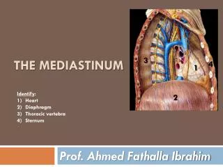
Mediastinum. Superior mediastinum. The superior mediastinum is posterior to the manubrium of the sternum and anterior to the bodies of the first four thoracic vertebrae. The superior mediastinum is continuous with the neck superiorly and with the inferior mediastinum inferiorly.

E N D
Superior mediastinum • The superior mediastinum is posterior to the manubrium of the sternum and anterior to the bodies of the first four thoracic vertebrae. • The superior mediastinum is continuous with the neck superiorly and with the inferior mediastinum inferiorly.
Major structures found in the superior mediastinum • Thymus • Right and left brachiocephalic veins • Left superior intercostal vein • Superior vena cava • Arch of the aorta with its three large branches • Trachea • Esophagus
Phrenicnerves • Vagusnerves • Left recurrent laryngeal branch of the left vagus nerve • Thoracic duct
Thymus • The thymus is the most anterior component of the superior mediastinum, lying immediately posterior to the manubrium of the sternum. It is an asymmetric, bilobedstructure. • The upper extent of the thymus can reach into the neck as high as the thyroid gland; a lower portion typically extends into the anterior mediastinum over the pericardial sac.
Right and left brachiocephalic veins • The left and right brachiocephalic veins are located immediately posterior to the thymus. • They form on each side at the junction between the internal jugular and subclavian veins • The left brachiocephalic vein crosses the midline and joins with the right brachiocephalic vein to form the superior vena cava
Left superior intercostal vein • The left superior intercostal vein receives the second, third and sometimes the fourth posterior intercostalveins. • Enters the left brachiocephalicvein.
Superior vena cava • The vertically oriented superior vena cava begins posterior to the lower edge of the right first costal cartilage, where the right and left brachiocephalic veins join, and terminates at the lower edge of the right third costal cartilage, where it joins the right atrium. • The superior vena cava receives the azygos vein immediately before entering the pericardial sac
Arch of aorta and its branches • Only the arch of the aorta is in the superior mediastinum. • It begins when the ascending aorta emerges from the pericardial sac and courses upward, backward, and to the left as it passes through the superior mediastinum, ending on the left side at vertebral level TIV/V.
Ligamentumarteriosum • The ligamentumarteriosum is also in the superior mediastinum and is important in embryonic circulation, when it is a patent vessel (the ductusarteriosus). • It connects the pulmonary trunk with the arch of aorta and allows blood to bypass the lungs during development
Trachea and esophagus • The trachea is a midline structure that is palpable in the jugular notch as it enters the superior mediastinum. • Posterior to it is the esophagus, which is immediately anterior to the vertebral column.
Nerves of the superior mediastinum • Vagus nerves :The vagus nerves [X] pass through the superior and posterior divisions of the mediastinum on their way to the abdominal cavity. • As they pass through the thorax, they provide parasympathetic innervation to the thoracic viscera
Left recurrent laryngeal nerve • The left vagus nerve also gives rise to the left recurrent laryngeal nerve, which arises from it at the inferior margin of the arch of aorta just lateral to the ligamentumarteriosum. • Entering a groove between the trachea and esophagus, the left recurrent laryngeal nerve continues superiorly to enter the neck and terminate in the larynx.
Thoracic duct in the superior mediastinum • The thoracic duct, which is the major lymphatic vessel in the body, passes through the posterior portion of the superior mediastinum.
Posterior mediastinum • The posterior mediastinum is posterior to the pericardial sac and diaphragm and anterior to the bodies of the mid and lower thoracic vertebrae
Major structures in the posterior mediastinum • Esophagus and its associated nerve plexus • Thoracic aorta and its branches • Azygossystem of veins • Thoracic duct and associated lymph nodes • Sympathetic trunks • Thoracic splanchnicnerves
Esophagus • Relationships to important structures in the posterior mediastinum • 1. Posterior to the esophagus, the thoracic duct is on the right side inferiorly, but crosses to the left more superiorly. • 2. Esophagus passes immediately posteriorly to the left atrium, separated from it only by pericardium
Thoracic aorta • The thoracic portion of the descending aorta (thoracic aorta) begins at the lower edge of vertebra TIV. • Branches of the thoracic aorta • 1. Pericardial branches • 2. Bronchial branches • 3. Esophageal branches • 4. Posterior intercostalarteries • 5. Superior phrenicarteries • 6. Subcostal artery
Azygos vein • The azygos vein arises opposite vertebra LI or LII at the junction between the right ascending lumbar vein and the right subcostalvein. • The azygos vein enters the thorax through the aortic hiatus of the diaphragm. • At approximately vertebral level TIV, it arches anteriorly, over the root of the right lung, to join the superior vena cava
Thoracic duct in the posterior mediastinum • The thoracic duct is the principal channel through which lymph from most of the body is returned to the venous system. • It begins as a confluence of lymph trunks in the abdomen, sometimes forming a saccular dilation referred to as the cisternachyli (chyle cistern), which drains the abdominal viscera and walls, pelvis, perineum, and lower limbs. • Thoracic duct empties into the junction of the left subclavian and left internal jugular veins.
Sympathetic trunks • Sympathetic trunks consists of two parallel cords punctuated by 11 or 12 ganglia. • Thoracic splanchnicnerves • 1. Greater splanchnicnerve • 2. Lesser splanchnicnerve • 3. Least splanchnic nerve (lowest splanchnic nerve)
Anterior mediastinum • The anterior mediastinum is posterior to the body of the sternum and anterior to the pericardial sac • CONTENTS: • Sternoparicardialligaments • Few lymph nodes • Branches of the internal thoracic vessels. • In infants and children, the anterior mediastinum contains the inferior part of the thymus.