Download
1 / 45
490 likes | 1.24k Views
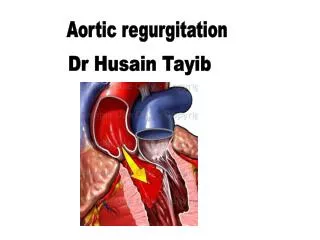
Clinical hemodynamic correlation in aortic regurgitation. Dr.Deepak Raju. Etiology . Aortic root disease Aortopathy Aortitis Age related aortic dilatation Valvular disease Calcific AS in older patients with AR Bicuspid aortic valve Cusp retraction or fibrosis c/c rheumatic

E N D
Clinical hemodynamic correlation in aortic regurgitation Dr.DeepakRaju
Etiology • Aortic root disease • Aortopathy • Aortitis • Age related aortic dilatation • Valvular disease • Calcific AS in older patients with AR • Bicuspid aortic valve • Cusp retraction or fibrosis • c/c rheumatic • Inflammatory • Cusp perforation/tears • Infective endocarditis • Trauma • Lack of cusp support • Dissection of aorta • VSD
Volume overload –compensatory mechanisms • LV EDV increases without increase in diastolic pressure due to increased compliance • LV preload reserve is maintained initially • Eccentric hypertrophy • Sarcomeres laid in series • Preload at sarcomere level is near normal • Normal contractile performance of each unit contributes to enhanced stroke volume
Increased afterload • Increased chamber volume,increased systolic pressure • Increased systolic wall stress and afterload • concentric LVH • Continued increase in chamber volume and afterload –matched by continued recruitment of preload reserve and compensatory hypertrophy
Decompensation • Afterload mismatch-reversible • Impaired LV contractility-irreversible
A/c AR-pathophysiology • Hemodynamically significant AR of sudden onset,into a LV not previously subjected to volume overload • Volume overload is poorly tolerated • Ventricular compliance is normal • LV operating on steep portion of diastolic P/V relation • End diastolic LV pressure markedly increased approaching aortic diastolic pressure
LV fails to increase stroke voume(not hypertrophied or dilated)-Decrease in COP • Increase in LVEDP causes rise in mean LA pressure and PCWP-pulmonary edema • Premature closure of MV –early crossover of pressures • Diastolic MR • Arterial BP- • fall in syst pr • Normal pulse pressure • Diastolic pressure maintained by reflex increase in SVR in failure
Hemodynamic assessment-c/c AR • Elevated Ao.syst pressure • Lowered Ao.diastolic pressure • modest rise of LV pressures in diastole • Premature closure of MV-when LV diastolic pressure exceeds LA pressure-common in a/c AR • Mean diastolic pressures rise with time and severity of leak-rise in mean LA &PCWP • Amplification of peak systolic pressure in peripheral arteries
Acute AR • Regurgitation into non compliant LV -diastolic rise of LV pressure&absence of A wave • LV diastolic pressure exceeds LA pressure-premature closure of MV • Aortic and LV pressures equalise in diastole and regurgitant flow &murmur ceases
Angiographic assessment • Mild(1+) • small amount of contrast • never fills chamber • cleared with each beat • Moderate(2+) • more contrast • faint opacification of entire chamber • Moderately severe(3+) • LV well opacified • equal in density with aorta • Severe(4+) • complete dense opacification of LV in one beat • LV more densely opacified than aorta
Clinical features • Asymptomatic phase longer • Dyspnoea most common symptom • Angina in 20% patients • Decreased perfusion-low aortic diastolic pressure • Increased myocardial oxygen demand • Associated coronary atherosclerosis • Osteal coronary invt.in syphilitic AR,takayasuarteritis • Palpitations- • awareness of forceful ventricular contraction • Ventricular arrhythmias in decompensated stage • Syncope-5 to 10%
Physical findings • Elevated systolic pressure • Low diastolic pressure • Peripheral signs of AR-large stroke volume in early systole with subsequent run off • Hill s sign-exaggeration of peripheral amplification • Carotid thrill or shudder-more common in AS,but also in AR • Displacement of apical impulse
S1-soft • Increased LVEDP-earlier closure of MV • Elevated diastolic pressure-less valve excursion • S2- • Soft A2 –valve structurally abnormal • Delayed A2-prolonged LV ejection time • P2 may be obscured by murmur • S3 – • in failure • S4- • Suggest decreased LV compliance&increased LVEDP • Long PR interval
Early diastolic murmur • high pitched in mild to moderate,pitch decreases as severity increases • Decrescendo-aortic LV pressure gradient tapers in diastole • Duration • correlates with severity in most cases • Some patients with severe AR can have shorter murmur due to high LVEDP • Murmur shorter in decompensation • Murmur in 3rd RICS louder than 3rd LICS-Harvey s sign-AR is due to disease process involving aortic root-rightward and superior displacement of dilated proximal aorta • Seagull murmur-eversion or perforation of a valve cusp
Systolic ejection murmur • Increased LV stroke volume • Abnormal Aortic valve
Austin Flint murmur • low pitched,mid or late diastolic murmur • Mechanism • AR jet pushing AML • Antegradetransmitral blood flow across a functionally narrowed MV • Diastolic MR • Low pitched components of AR murmur heard best at apex • Severe AR-reg. fraction>50% • Severity of AR and AFM • Mild-absent • Moderate-may be present in late diastole • Severe-earlier in timing,extend into presystole • Very severe AR-premature closure of MV-absent presystolic component
A/c AR • Rapid onset of symptoms – • rapid rise of LA pressure • abrupt reduction of COP • BP- • Systolic pressure normal or slight fall • elevated dia.pressure • narrrow pulse pressure • Acute rt heart failure can occur-elevated JVP
Soft S1 • Soft A2,loud P2 • LV S3-rapid early diastolic filling • Absent LVS4 • EDM • Short -rapid diastolic equilibration of aortic and LV pressures in diastole • Low or medium pitch- • Low gradient • a/w CCF • Austin Flint murmur presystolic component absent
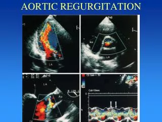
Echocardiography • Increased LV End Diastolic Dimensions ,near normal end systolic dimensions and increased contractility-compensated phase • Increase in end systolic dimensions and depressed contractility-decompensation • M-Mode of MV • Diastolic fluttering of AML in c/c AR • Early closure of MV in a/c AR • M-mode of AV • Diastolic non coaptation,diastolic fluttering in c/c AR • Premature opening of AV in a/c AR
SEVERITY • 1.Regurgitantjet width/LVOT diameter ratio greater than or equal to 60 percent • 2.Vena contracta greater than 6 mm • 3.Regurgitantjet area/LVOT area ratio greater than or equal to 60 percent • 4. Aortic regurgitation pressure half-time less than or equal to 250 ms • 5.Holodiastolicflow reversal in the descending thoracic or abdominal aorta • 6.Regurgitant volume greater than or equal to 60 mL • 7.Regurgitant fraction greater than or equal to 50 percent • 8.Effective regurgitant orifice greater than or equal to 0.30cm2 • 9.Restrictive mitral flow pattern (usually in acute setting)
1.Regurgitant jet width/LVOT diameter ratio greater than or equal to 60 percent
3.Regurgitant jet area/LVOT area ratio greater than or equal to 60 percent
CW doppler of AR jet • PHT and deceleration slope in severity assessment • AR PHT <250 ms or deceleration slope >400 cm/s • Overestimates AR in patients with high LVEDP due to other causes • Depends on LV compliance • PHT limited by technical factors-recording of peak velocity
5.Holodiastolic flow reversal in the descending thoracic or abdominal aorta
Quantitative measurements • Regurgitant volume=SV lvot-SV mv/pv • Regurgitant fraction=reg volume/total stroke volume • ERO=reg.volume/VTI reg. • Advantage • Measures independent of loading conditions or LV compliance • Limitations • Small errors in annulus size measurements-large error in volume calculations • Accuracy reduced outflow tract obstruction,shunts • Forward stroke volume estimation affected by MR/PR