Download

1 / 81
881 likes | 2k Views
Clinical hemodynamic correlation in mitral stenosis. Dr.Deepak Raju. Grading of severity in MS. Normal CSA of mitral valve – 4 to 5 cm2 No significant gradient across normal mitral valve during diastolic flow Progressive narrowing of mitral orifice results in

E N D
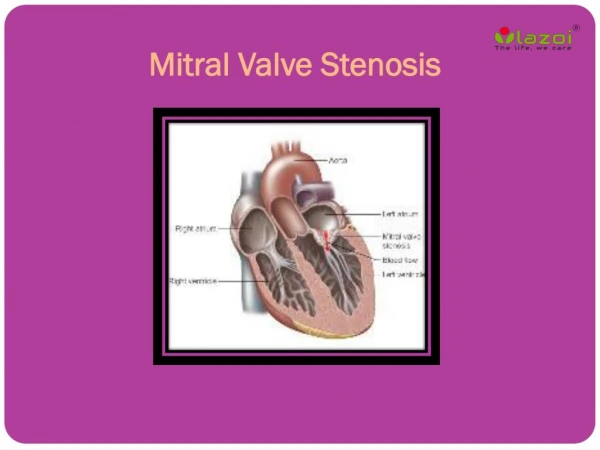
Clinical hemodynamic correlation in mitral stenosis Dr.DeepakRaju
Normal CSA of mitral valve – 4 to 5 cm2 • No significant gradient across normal mitral valve during diastolic flow • Progressive narrowing of mitral orifice results in • Pressure gradient b/w LA and LV • Left ventricular end diastolic pressure remaining at 5 mm Hg,LA mean pressure rises gradually • Reaches around 25 mmHg when MVA around 1 cm2 • Reduction of blood flow across mitral valve • COP 3.0 L/min /m2 falls to around 2.5 L/min /m2 at MVA 1 cm2 • Dependence of LV filling on LA pressure • Elevation of LA mean pressure-pulmonary venous hypertension
Factors affecting transmitral gradient • √mean grad∞ COP/DFP*MVA • Factors ↑ grad • ↑ COP • Exertion ,emotion,high output states • ↓ DFP • Increase HR • ↓ MVA • Progression of disease • thrombus
Factors decreasing gradient • ↓ COP • Second stenosis • RV failure • ↑ DFP • Slow HR • ↑ MVA
↑pul venous pressure • Transudation of fluid into interstitium • Initially lymphatic drainage increases to drain excess fluid-fails as venous pressure increases • Transudate decrease lung compliance-increase work of breathing • Bronchospasm,Alveolarhypoxia,vasoconstriction • Symptoms-dyspnoea,orthopnoea,PND
a/c pulmonary edema • PCWP exceeds tissue oncotic pressure of 25 mmHg&lymphatics unable to decompress the transudate • Gradual in a tight MS or abrupt appearance in a moderate to severe MS a/w ↑HR or ↑ transvalvular flow • Onset of AF • tachycardia • Fluid overload • Pregnancy • High output states
Hemoptysis • Pulmonary apoplexy • Sudden,profuse,bright red • Sudden increase in pulmonary venous pressure&rupture of bronchial vein collaterals • Pink frothy sputum of pulmonary edema • Blood stained sputum of PND • Blood streaked sputum a/w bronchitis • Pulmonary infarction
Winter bronchitis • Pulmonary venous hypertension-c/c passive congestion of lung-bronchial hyperemia • Hypersecretion of seromucinous glands –excessive mucus production • Symptoms of bronchitis
Effects of c/c elevation of pul venous pressure • Increase in lymphatic drainage • Engorged systemic bronchial veins • Pulmonary arterial hypertension
Pulmonary HTN • Devt of pulmonary hypertension • Passive • Active • Organic obliterative changes • Passive pulmonary HTN • Obligatory increase in response to ↑PCWP to maintain gradient of 10 to 12 across pulvasc bed(PA mean-LA mean) • Active pulmonary HTN • PA mean pressure –LA mean pressure >10 to 12
Cause of reactive pul HTN • Wood-pulmonary vasoconstriction • Doyle-↑pul venous pressure prominent in the lower lobes,produce reflex arterial constriction • Heath &Harris-↑ PA pressure causes reflex arteriolar constriction
Jordan- • ↑pul venous pressure-transudation of fluid • causes thickening and fibrosis of alveolar walls • hypoventilation of lower lobes-hypoxemia in lower lobe vessels • Sensed by chemoreceptors in pulmonary veins • Pulmonary arteriolar vasoconstriction in regions supplying these alveoli • Lower lobe perfusion decreases • This process eventually involve middle and upper lobe
Anatomical changes in the pulmonary arterioles • Medial hypertrophy • Intimal proliferation • Fibrosis • Decrease in CSA of pulmonary vascular bed • Increase PVR
Sequlae of reactive pul HTN • RV hypertrophy • Functional TR • RV failure
Symptoms and hemodynamic correlation • Precapillary block • Low cardiac output • Right ventricular hypertrophy • RV dysfunction • Postcapillary block • Left sided failure
Stage 1 • Asymptomatic at rest • Stage 2 • Symptomatic due to elevated LA pressure • Normal pulmonary vasc resistance • Stage 3 • Increased pulmonary vascular resistance • Relatively asymptomatic OR symptoms of low COP • Stage 4 • Both stenoses severe • Extreme elevation of PVR-RV failure
Elevated precapillary resistance protects against devt of pulmonary congestion at cost of a reduced COP • Severe pulmonary HTN leads to right sided failure
Exercise hemodynamics-2 types of response • Normal COP&hightransvalvular gradient-symptomatic due to pulmonary congestion • Reduced COP &low gradient-symptoms of low COP • Severe MS-combination of low output and pulmonary congestion symptoms
Role of LA compliance • Non compliant LA • Severe elevation of LA pressure and congestive symptoms • Dilated compliant LA • Decompress LA pressure • PHT =11 .6*Cn*√ MPG/(Cc*MVA) • Cn-net compliance • Thomas JD (circulation 1988) • Post BMV • Reduction of LV compliance <improvement in LA compliance • Net compliance increases-overestimate PHT • MVA underestimated
Impact of AF in MS • ↑HR,↓DFP-elevates transmitral gradient • Loss of atrial contribution to LV filling • Normal contribution of LA contraction to LV filling 15% • In MS,increasesupto 25-30% • Lost in AF • Loss of A wave in M-mode echo and in LA pressure tracing
Physical findings and correlation • Pulse-normal or low volume in ↓ COP • JVP- • mean elevated in RV failure • prominent a wave in PAH in SR • Absent a wave in AF • Palpation • Apical impulse • Inconspicous LV • Tapping S1 • RV apex in exreme RVH • LPH in RVH • Palpable P2
Loud S1 • Mitral valve closes at a higher Dp/dt of LV • In MS closure of mitral valve is late due to elevated LA pressure • LA –LV pressure crossover occurs after LV pressure has begun to rise • Rapidity of pressure rise in LV contributes to closing of MV to produce a loud S1 • Wide closing excursion of leaflets • Persistent LA-LV gradient in late diastole keeps valve open and at a lower position into late diastole • Increased distance that traversed during closing motion contributes to loud S1 • Quality of valve tissue may affect amplitude of sound • The diseased MV apparatus may resonate with a higher amplitude than normal tissue
Soft S1 &decreased intensity of OS in severe MS • MV Calcification especially AML • Severe PAH-reduced COP • CCF-reduced COP • Large RV • AS-reduced LV compliance • AR • Predominant MR • LV dysfunction
Q-S1 interval • Prolongation of Q-S1 interval • As LA pressure rises,LA-LV pressure crossover occurs later • Well’s index- • Q-S1 interval-A2 OS interval expressed in units of 0.01 sec • >2 unit correlate with MVA <1.2 cm2
S2 • Loud P2 • Narrow split as PAH increases • Reduced compliance and earlier closure of pulmonary valve • RVS4 • LVS3 rules out significant MS
A2-OS interval • OS- • Sudden tensing of valve leaflets after the valve cusps have completed their opening excursion • Movement of mitral dome into LV suddenly stops • Follows LA LV pressure crossover in early diastole by 20-40 ms • A2 OS interval ranges from 40 -120 ms • As LA pressure rises,the crossover of LA and LV pressure occurs earlier –MV opening motion begins earlier- A2 OS interval shortens • Narrow A2 OS interval <80 ms-severe MS
Short A2 OS interval • Severe MS • Tachycardia • Associated MR-Higher LA pressure –MV open earlier • Long A2-OS interval in severe MS • Factors that affect MV opening –AR,MV calcification • Factors that decrease LV compliance-AS,systHTN,old age • Decreased rate of pressure decline in LV during IVRT as in LV dysfunction • Due to low LA pressure in a large compliant LA • In AF-shorter cycle length-LA pressure remains elevated-A2 OS narrows
Diastolic murmur of MS • Two components- • early diastolic component that begins with the opening snap,whenisovolumic LV pressure falls below LA pressure • Late diastolic component • Increase in LA-LV pressure gradient due to atrial systole • Persistence of LA-LV gradient upto late diastole in severe MS • closing excursion of mitral valve produces a decreasing orifice area • velocity of flow increases as valve orifice narrows • this cause turbulence to produce presystolic murmur
Duration of murmur correlates with severity • Murmur persists as long as transmitral gradient>3 mmHg • Mild MS- • murmur in early diastole • or in presystole with crescendo pattern • or both murmurs present with a gap b/w components • Moderate to severe MS- • murmur starts with OS and persists upto S1
Presystolic accentuation of murmur • Atrial contraction in patients in sinus rhythm • Reduction in mitral valve orifice by LV contraction • Increase velocity of flow as long as there is a pressure gradient LA-LV • Persistence of presystolic accentuation in AF in severe MS
Factors that decrease intensity of diastolic murmur of MS • Low flow states • Severe MS • Severe PAH • CCF • AF with rapid ventricular rate • Associated cardiac lesions • Aortic stenosis-LVH,decreased compliance-decreased opening motion of mitral valve • Aortic regurgitation • ASD • PHT with marked RV enlargement
Characteristics of mitral valve • Extensive calcification • Others • Apex formed by RV • Inability to localise apex • Obesity • Muscular chest • COPD
Factors increasing intensity of murmur • a/w MR-increased volume of LA blood-increased transvalvular flow • Tachycardia
Calculation of MVA • Toricelli’s law • F=AVCc • A=F/V Cc • F-Flow rate,A-orifice area,V-velocity of flow • Cc-coefficient of orifice contraction • Gradient and velocity of flow related by • V 2=Cv2*2 g h • G=gravitational constant,h=pressure gradient • Cv=Coefficient of Velocity • V=Cv*√2 g h • MVA=F/Cv*Cc* √2 g h =F/C*44.3*√h
Flow • Total cardiac output divided by time in seconds during which flow occurs across the valve • F=COP/DFP*HR
Steps • Average gradient=area(mm2)/length of diastole(mm) • Mean gradient=average gr * scale • Average diastolic period=length of DFP(mm)/paper speed(mm/s) • HR(bt/min),COP(ml/min) • MVA=cardiac output/HR×average diastolic period÷37.7×√mean gradient